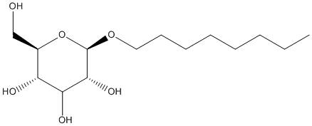
6 structures(e.g. : 7OTS, 6I8W, 1K8Q... more)(less)7OTS: Crystal structure of human Monoacylglycerol Lipase ABHD6 in complex with oleic acid and octyl glucoside, 6I8W: Crystal structure of a membrane phospholipase A from Pseudomonas aeruginosa, a novel bacterial virulence factor, 1K8Q: Dog gastric lipase complex with a phosphonate inhibitor, 1TCB: Candida antarctica triacylglycerol lipase form B 2.1 Angst, 1TCC: Candida antarctica triacylglycerol lipase form B 2.5 Angst, 1VE6: Crystal structure of an acylpeptide hydrolase/esterase from Aeropyrum pernix K1
Lipase inhibitors are the main anti-obesity drugs prescribed these days, but the complexity of their mechanism of action is making it difficult to develop new molecules for this purpose. The efficacy of these drugs is known to depend closely on the physico-chemistry of the lipid-water interfaces involved and on the unconventional behavior of the lipases which are their target enzymes. The lipolysis reaction which occurs at an oil-water interface involves complex equilibria between adsorption-desorption processes, conformational changes and catalytic mechanisms. In this context, surfactants can induce significant changes in the partitioning of the enzyme and the inhibitor between the water phase and lipid-water interfaces. Surfactants can be found at the oil-water interface where they compete with lipases for adsorption, but also in solution in the form of micellar aggregates and monomers that may interact with hydrophobic parts of lipases in solution. These various interactions, combined with the emulsification and dispersion of insoluble substrates and inhibitors, can either promote or decrease the activity and the inhibition of lipases. Here, we review some examples of the various effects of surfactants on lipase structure, activity and inhibition, which show how complex the various equilibria involved in the lipolysis reaction tend to be.
Acylpeptide hydrolases (APH; also known as acylamino acid releasing enzyme) catalyze the removal of an N-acylated amino acid from blocked peptides. The crystal structure of an APH from the thermophilic archaeon Aeropyrum pernix K1 to 2.1 A resolution confirms it to be a member of the prolyl oligopeptidase family of serine proteases. The structure of apAPH is a symmetric homodimer with each subunit comprised of two domains. The N-terminal domain is a regular seven-bladed beta-propeller, while the C-terminal domain has a canonical alpha/beta hydrolase fold and includes the active site and a conserved Ser445-Asp524-His556 catalytic triad. The complex structure of apAPH with an organophosphorus substrate, p-nitrophenyl phosphate, has also been determined. The complex structure unambiguously maps out the substrate binding pocket and provides a basis for substrate recognition by apAPH. A conserved mechanism for protein degradation from archaea to mammals is suggested by the structural features of apAPH.
        
Title: The sequence, crystal structure determination and refinement of two crystal forms of lipase B from Candida antarctica Uppenberg J, Hansen MT, Patkar S, Jones TA Ref: Structure, 2:293, 1994 : PubMed
BACKGROUND: Lipases constitute a family of enzymes that hydrolyze triglycerides. They occur in many organisms and display a wide variety of substrate specificities. In recent years, much progress has been made towards explaining the mechanism of these enzymes and their ability to hydrolyze their substrates at an oil-water interface. RESULTS: We have determined the DNA and amino acid sequences for lipase B from the yeast Candida antarctica. The primary sequence has no significant homology to any other known lipase and deviates from the consensus sequence around the active site serine that is found in other lipases. We have determined the crystal structure of this enzyme using multiple isomorphous replacement methods for two crystal forms. Models for the orthorhombic and monoclinic crystal forms of the enzyme have been refined to 1.55 A and 2.1 A resolution, respectively. Lipase B is an alpha/beta type protein that has many features in common with previously determined lipase structures and other related enzymes. In the monoclinic crystal form, lipid-like molecules, most likely beta-octyl glucoside, can be seen close to the active site. The behaviour of these lipid molecules in the crystal structure has been studied at different pH values. CONCLUSION: The structure of Candida antarctica lipase B shows that the enzyme has a Ser-His-Asp catalytic triad in its active site. The structure appears to be in an 'open' conformation with a rather restricted entrance to the active site. We believe that this accounts for the substrate specificity and high degree of stereospecificity of this lipase.
        
2 lessTitle: Pseudomonas aeruginosa esterase PA2949, a bacterial homolog of the human membrane esterase ABHD6: expression, purification and crystallization Bleffert F, Granzin J, Gohlke H, Batra-Safferling R, Jaeger KE, Kovacic F Ref: Acta Crystallographica F Struct Biol Commun, 75:270, 2019 : PubMed
The human membrane-bound alpha/beta-hydrolase domain 6 (ABHD6) protein modulates endocannabinoid signaling, which controls appetite, pain and learning, as well as being linked to Alzheimer's and Parkinson's diseases, through the degradation of the key lipid messenger 2-arachidonylglycerol (2-AG). This makes ABHD6 an attractive therapeutic target that lacks structural information. In order to better understand the molecular mechanism of 2-AG-hydrolyzing enzymes, the PA2949 protein from Pseudomonas aeruginosa, which has 49% sequence similarity to the ABHD6 protein, was cloned, overexpressed, purified and crystallized. Overexpression of PA2949 in the homologous host yielded the membrane-bound enzyme, which was purified in milligram amounts. Besides their sequence similarity, the enzymes both show specificity for the hydrolysis of 2-AG and esters of medium-length fatty acids. PA2949 in the presence of n-octyl beta-D-glucoside showed a higher activity and stability at room temperature than those previously reported for PA2949 overexpressed and purified from Escherichia coli. A suitable expression host and stabilizing detergent were crucial for obtaining crystals, which belonged to the tetragonal space group I4122 and diffracted to a resolution of 2.54 A. This study provides hints on the functional similarity of ABHD6-like proteins in prokaryotes and eukaryotes, and might guide the structural study of these difficult-to-crystallize proteins.
Lipase inhibitors are the main anti-obesity drugs prescribed these days, but the complexity of their mechanism of action is making it difficult to develop new molecules for this purpose. The efficacy of these drugs is known to depend closely on the physico-chemistry of the lipid-water interfaces involved and on the unconventional behavior of the lipases which are their target enzymes. The lipolysis reaction which occurs at an oil-water interface involves complex equilibria between adsorption-desorption processes, conformational changes and catalytic mechanisms. In this context, surfactants can induce significant changes in the partitioning of the enzyme and the inhibitor between the water phase and lipid-water interfaces. Surfactants can be found at the oil-water interface where they compete with lipases for adsorption, but also in solution in the form of micellar aggregates and monomers that may interact with hydrophobic parts of lipases in solution. These various interactions, combined with the emulsification and dispersion of insoluble substrates and inhibitors, can either promote or decrease the activity and the inhibition of lipases. Here, we review some examples of the various effects of surfactants on lipase structure, activity and inhibition, which show how complex the various equilibria involved in the lipolysis reaction tend to be.
Acylpeptide hydrolases (APH; also known as acylamino acid releasing enzyme) catalyze the removal of an N-acylated amino acid from blocked peptides. The crystal structure of an APH from the thermophilic archaeon Aeropyrum pernix K1 to 2.1 A resolution confirms it to be a member of the prolyl oligopeptidase family of serine proteases. The structure of apAPH is a symmetric homodimer with each subunit comprised of two domains. The N-terminal domain is a regular seven-bladed beta-propeller, while the C-terminal domain has a canonical alpha/beta hydrolase fold and includes the active site and a conserved Ser445-Asp524-His556 catalytic triad. The complex structure of apAPH with an organophosphorus substrate, p-nitrophenyl phosphate, has also been determined. The complex structure unambiguously maps out the substrate binding pocket and provides a basis for substrate recognition by apAPH. A conserved mechanism for protein degradation from archaea to mammals is suggested by the structural features of apAPH.
Fat digestion in humans and some mammals such as dogs requires the successive intervention of two lipases: gastric lipase, which is stable and active despite the highly acidic stomach environment, followed by the classical pancreatic lipase secreted into the duodenum. We previously solved the structure of recombinant human gastric lipase (HGL) at 3.0-A resolution in its closed form; this was the first structure to be described within the mammalian acid lipase family. Here we report on the open structure of the recombinant dog gastric lipase (r-DGL) at 2.7-A resolution in complex with the undecyl-butyl (C11Y4) phosphonate inhibitor. HGL and r-DGL show 85.7% amino acid sequence identity, which makes it relevant to compare the forms from two different species. The open r-DGL structure confirms the previous description of the HGL catalytic triad (Ser(153), His(353), and Asp(324)) with the catalytic serine buried and an oxyanion hole (NH groups of Gln(154) and Leu(67)). In r-DGL, the binding of the C11Y4 phosphonate inhibitor induces part of the cap domain, the lid, to roll over the enzyme surface and to expose a catalytic crevice measuring approximately 20 x 20 x 7 A(3). The C11Y4 phosphonate fits into this crevice, and a molecule of beta-octyl glucoside fills up the crevice. The C11Y4 phosphonate inhibitor and the detergent molecule suggest a possible binding mode for the natural substrates, the triglyceride molecules.
        
Title: The sequence, crystal structure determination and refinement of two crystal forms of lipase B from Candida antarctica Uppenberg J, Hansen MT, Patkar S, Jones TA Ref: Structure, 2:293, 1994 : PubMed
BACKGROUND: Lipases constitute a family of enzymes that hydrolyze triglycerides. They occur in many organisms and display a wide variety of substrate specificities. In recent years, much progress has been made towards explaining the mechanism of these enzymes and their ability to hydrolyze their substrates at an oil-water interface. RESULTS: We have determined the DNA and amino acid sequences for lipase B from the yeast Candida antarctica. The primary sequence has no significant homology to any other known lipase and deviates from the consensus sequence around the active site serine that is found in other lipases. We have determined the crystal structure of this enzyme using multiple isomorphous replacement methods for two crystal forms. Models for the orthorhombic and monoclinic crystal forms of the enzyme have been refined to 1.55 A and 2.1 A resolution, respectively. Lipase B is an alpha/beta type protein that has many features in common with previously determined lipase structures and other related enzymes. In the monoclinic crystal form, lipid-like molecules, most likely beta-octyl glucoside, can be seen close to the active site. The behaviour of these lipid molecules in the crystal structure has been studied at different pH values. CONCLUSION: The structure of Candida antarctica lipase B shows that the enzyme has a Ser-His-Asp catalytic triad in its active site. The structure appears to be in an 'open' conformation with a rather restricted entrance to the active site. We believe that this accounts for the substrate specificity and high degree of stereospecificity of this lipase.