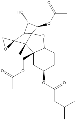
T-2 toxin is a trichothecene mycotoxin produced by fungi of the genus Fusarium. It is a common contaminant in food and feedstuffs of cereal origin Elicits inflammatory reaction in animals and has teratogenic effects; (cardiotoxic neurotoxic, apoptosis inducer, DNA synthesis inhibitor) hydrolyzed to HT-2 toxin by carboxylesterases (Jia 2022). HT-2 (with one ester left) is also toxic. T-2 triol (with two esters hydrolyzed) is less toxic. T-2 triol is the end product of hydrolysis by 9bact-estC55.8n1/IS12 from Chloroflexi bacterium. HT-2 is the end product of IS10. Among FamilyVIII beta lactamses some hydrolyze T-2 (Cea-Rama_2022) and some do not ISL11 (Distaso 2023)
BACKGROUND: T-2 toxin is recognized as one of the high-risk environmental factors for etiology and pathogenesis of Kashin-Beck disease (KBD). Previous evidence indicates decreased serum fluorine level in KBD patients. However, whether fluoride could regulate carboxylesterase 1 (CES1)-mediated T-2 toxin hydrolysis and alter its chondrocyte toxicity remains largely unknown. METHODS: In this study, in vitro hydrolytic kinetics were explored using recombinant human CES1. HPLC-MS/MS was used to quantitative determination of hydrolytic metabolites of T-2 toxin. HepG2 cells were treated with different concentration of sodium fluoride (NaF). qRT-PCR and western blot analysis were used to compare the mRNA and protein expression levels of CES1. C28/I2 cells were treated with T-2 toxin, HT-2 toxin, and neosolaniol (NEO), and then cell viability was determined by MTT assay, cell apoptosis was determined by Annexin V-FITC/PI, Hoechst 33258 staining, and cleaved caspase-3, and cell cycle was monitored by flow cytometry assay, CKD4 and CDK6. RESULTS: We identified that recombinant human CES1 was involved in T-2 toxin hydrolysis to generate HT-2 toxin, but not NEO, and NaF repressed the formation of HT-2 toxin. Both mRNA and protein expression of CES1 were significantly down-regulated in a dose-dependent manner after NaF treatment in HepG2 cells. Moreover, we evaluated the chondrocyte toxicity of T-2 toxin and its hydrolytic metabolites. Results showed that T-2 toxin induced strongest cell apoptosis, followed by HT-2 toxin and NEO. The decreased the proportion of cells in G0/G1 phase was observed with the descending order of T-2 toxin, HT-2 toxin, and NEO. CONCLUSIONS: This study reveals that CES1 is responsible for the hydrolysis of T-2 toxin, and that fluoride impairs CES1-mediated T-2 toxin detoxification to increase its chondrocyte toxicity. This study provides novel insight into understanding the relationship between fluoride and T-2 toxin in the etiology of KBD.
T-2 toxin (T-2) is a type-A trichothecene produced by Fusarium that causes toxicity to animals. T-2 contamination of grain-based aquatic feed is a concern for the industries related to edible aquatic crustacean species such as the shrimp industry because it can lead to serious food safety issues. T-2, its metabolites, and selected phase I (EROD, CarE) and phase II (GST, UGT, SULT) detoxification enzymes in hemolymph and tissues were monitored at 0, 5, 10 15, 30, 45, and 60 min following T-2 intramuscular administration (3 mg/kg bw) in shrimp (Litopenaeus vannamei). Marked increases of EROD activity in hepatopancreas and CarE activity in hemolymph, gill, hepatopancreas and intestine were observed followed by increases in phase II enzymes (GST, UGT, SULT) in hepatopancreas, hemolymph, intestine and gill, which remained elevated for an extended period. Time-dependent decrease in shrimp tissue T-2 concentration was observed. HT-2 increased up to 15 min. Most other T-2 metabolites were detected but not T-2 tetraol. Enzyme responses on exposure to T-2 were tissue-specific and time-dependent. Detection results indicated that HT-2 may not be the only important metabolite in aquatic crustacean species. Further investigation into T-2 metabolite toxicity is needed to fully understand the food safety issues related to T-2.
        
Title: The roles of carboxylesterase and CYP isozymes on the in vitro metabolism of T-2 toxin Lin NN, Chen J, Xu B, Wei X, Guo L, Xie JW Ref: Mil Med Res, 2:13, 2015 : PubMed
BACKGROUND: T-2 toxin poses a great threat to human health because it has the highest toxicity of the currently known trichothecene mycotoxins. To understand the in vivo toxicity and transformation mechanism of T-2 toxin, we investigated the role of one kind of principal phase I drug-metabolizing enzymes (cytochrome P450 [CYP450] enzymes) on the metabolism of T-2 toxin, which are crucial to the metabolism of endogenous substances and xenobiotics. We also investigated carboxylesterase, which also plays an important role in the metabolism of toxic substances. METHODS: A chemical inhibition method and a recombinant method were employed to investigate the metabolism of the T-2 toxin by the CYP450 enzymes, and a chemical inhibition method was used to study carboxylesterase metabolism. Samples incubated with human liver microsomes were analyzed by high performance liquid chromatography-triple quadrupole mass spectrometry (HPLC- QqQ MS) after a simple pretreatment. RESULTS: In the presence of a carboxylesterase inhibitor, only 20 % T-2 toxin was metabolized. When CYP enzyme inhibitors and a carboxylesterase inhibitor were both present, only 3 % of the T-2 toxin was metabolized. The contributions of the CYP450 enzyme family to T-2 toxin metabolism followed the descending order CYP3A4, CYP2E1, CYP1A2, CYP2B6 or CYP2D6 or CYP2C19. CONCLUSION: Carboxylesterase and CYP450 enzymes are of great importance in T-2 toxin metabolism, in which carboxylesterase is predominant and CYP450 has a subordinate role. CYP3A4 is the principal member of the CYP450 enzyme family responsible for T-2 toxin metabolism. The primary metabolite produced by carboxylesterase is HT-2, and the main metabolite produced by CYP 3A4 is 3'-OH T-2. The different metabolites show different toxicities. Our results will provide useful data concerning the toxic mechanism, the safety evaluation, and the health risk assessment of T-2 toxin.
Hydrothermal vents are geographically widespread and host microorganisms with robust enzymes useful in various industrial applications. We examined microbial communities and carboxylesterases of two terrestrial hydrothermal vents of the volcanic island of Ischia (Italy) predominantly composed of Firmicutes, Proteobacteria, and Bacteroidota. High-temperature enrichment cultures with the polyester plastics polyhydroxybutyrate and polylactic acid (PLA) resulted in an increase of Thermus and Geobacillus species and to some extent Fontimonas and Schleiferia species. The screening at 37 to 70 degreesC of metagenomic fosmid libraries from above enrichment cultures identified three hydrolases (IS10, IS11, and IS12), all derived from yet-uncultured Chloroflexota and showing low sequence identity (33 to 56%) to characterized enzymes. Enzymes expressed in Escherichia coli exhibited maximal esterase activity at 70 to 90 degreesC, with IS11 showing the highest thermostability (90% activity after 20-min incubation at 80 degreesC). IS10 and IS12 were highly substrate promiscuous and hydrolyzed all 51 monoester substrates tested. Enzymes were active with PLA, polyethylene terephthalate model substrate, and mycotoxin T-2 (IS12). IS10 and IS12 had a classical alpha/beta-hydrolase core domain with a serine hydrolase catalytic triad (Ser155, His280, and Asp250) in their hydrophobic active sites. The crystal structure of IS11 resolved at 2.92 A revealed the presence of a N-terminal beta-lactamase-like domain and C-terminal lipocalin domain. The catalytic cleft of IS11 included catalytic Ser68, Lys71, Tyr160, and Asn162, whereas the lipocalin domain enclosed the catalytic cleft like a lid and contributed to substrate binding. Our study identified novel thermotolerant carboxylesterases with a broad substrate range, including polyesters and mycotoxins, for potential applications in biotechnology. IMPORTANCE High-temperature-active microbial enzymes are important biocatalysts for many industrial applications, including recycling of synthetic and biobased polyesters increasingly used in textiles, fibers, coatings and adhesives. Here, we identified three novel thermotolerant carboxylesterases (IS10, IS11, and IS12) from high-temperature enrichment cultures from Ischia hydrothermal vents and incubated with biobased polymers. The identified metagenomic enzymes originated from uncultured Chloroflexota and showed low sequence similarity to known carboxylesterases. Active sites of IS10 and IS12 had the largest effective volumes among the characterized prokaryotic carboxylesterases and exhibited high substrate promiscuity, including hydrolysis of polyesters and mycotoxin T-2 (IS12). Though less promiscuous than IS10 and IS12, IS11 had a higher thermostability with a high temperature optimum (80 to 90 degreesC) for activity and hydrolyzed polyesters, and its crystal structure revealed an unusual lipocalin domain likely involved in substrate binding. The polyesterase activity of these enzymes makes them attractive candidates for further optimization and potential application in plastics recycling.
Family VIII esterases present similarities to class C beta-lactamases, which show nucleophilic serines located at the S-X-X-K motif instead of the G-X-S-X-G or G-D-S-(L) motif shown by other carboxylesterase families. Here, we report the crystal structure of a novel family VIII (subfamily VIII. I) esterase (EH(7) ; denaturing temperature, 52.6+/-0.3 degreesC; pH optimum 7.0-9.0) to deepen its broad substrate range. Indeed, the analysis of the substrate specificity revealed its capacity to hydrolyse nitrocefin as a model chromogenic cephalosporin substrate (40.4 +/- 11.4 units/g), as well as a large battery of 66 structurally different esters (up to 1730 min(-1) ), including bis(2-hydroxyethyl)-terephthalate (241.7 +/- 8.5 units/g) and the mycotoxin T-2 (1220 +/- 52 units/g). It also showed acyltransferase activity through the synthesis of benzyl 3-oxobutanoate (40.4 +/- 11.4 units/g) from benzyl alcohol and vinyl acetoacetate. Such a broad substrate scope is rare among family VIII esterases and lipolytic enzymes. Structural analyses of free and substrate-bound forms of this homo-octamer esterase suggest that EH(7) presents a more opened and exposed S1 site having no steric hindrance for the entrance of substrates to the active site, more flexible R1, R2 and R3 regions allowing the binding of a wide spectrum of substrates into the active site, as well as small residues in the conserved motif Y-X-X containing the catalytic Tyr enabling the entrance of large substrates. These unique structural elements in combination with docking experiments allowed us to gain valuable insights into the substrate specificity of this esterase and possible others belonging to family VIII.
BACKGROUND: T-2 toxin is recognized as one of the high-risk environmental factors for etiology and pathogenesis of Kashin-Beck disease (KBD). Previous evidence indicates decreased serum fluorine level in KBD patients. However, whether fluoride could regulate carboxylesterase 1 (CES1)-mediated T-2 toxin hydrolysis and alter its chondrocyte toxicity remains largely unknown. METHODS: In this study, in vitro hydrolytic kinetics were explored using recombinant human CES1. HPLC-MS/MS was used to quantitative determination of hydrolytic metabolites of T-2 toxin. HepG2 cells were treated with different concentration of sodium fluoride (NaF). qRT-PCR and western blot analysis were used to compare the mRNA and protein expression levels of CES1. C28/I2 cells were treated with T-2 toxin, HT-2 toxin, and neosolaniol (NEO), and then cell viability was determined by MTT assay, cell apoptosis was determined by Annexin V-FITC/PI, Hoechst 33258 staining, and cleaved caspase-3, and cell cycle was monitored by flow cytometry assay, CKD4 and CDK6. RESULTS: We identified that recombinant human CES1 was involved in T-2 toxin hydrolysis to generate HT-2 toxin, but not NEO, and NaF repressed the formation of HT-2 toxin. Both mRNA and protein expression of CES1 were significantly down-regulated in a dose-dependent manner after NaF treatment in HepG2 cells. Moreover, we evaluated the chondrocyte toxicity of T-2 toxin and its hydrolytic metabolites. Results showed that T-2 toxin induced strongest cell apoptosis, followed by HT-2 toxin and NEO. The decreased the proportion of cells in G0/G1 phase was observed with the descending order of T-2 toxin, HT-2 toxin, and NEO. CONCLUSIONS: This study reveals that CES1 is responsible for the hydrolysis of T-2 toxin, and that fluoride impairs CES1-mediated T-2 toxin detoxification to increase its chondrocyte toxicity. This study provides novel insight into understanding the relationship between fluoride and T-2 toxin in the etiology of KBD.
T-2 toxin is the most toxic trichothecene mycotoxin, and it exerts potent toxic effects, including immunotoxicity, neurotoxicity, and reproductive toxicity. Recently, several novel metabolites, including 3',4'-dihydroxy-T-2 toxin and 4',4'-dihydroxy-T-2 toxin, have been uncovered. The enzymes CYP3A4 and carboxylesterase contribute to T-2 toxin metabolism, with 3'-hydroxy-T-2 toxin and HT-2 toxin as the corresponding primary products. Modified forms of T-2 toxin, including T-2-3-glucoside, exert their immunotoxic effects by signaling through JAK/STAT but not MAPK. T-2-3-glucoside results from hydrolyzation of the corresponding parent mycotoxin and other metabolites by the intestinal microbiota, which leads to enhanced toxicity. Increasing evidence has shown that autophagy, hypoxia-inducible factors, and exosomes are involved in T-2 toxin-induced immunotoxicity. Autophagy promotes the immunosuppression induced by T-2 toxin, and a complex crosstalk between apoptosis and autophagy exists. Very recently, "immune evasion" activity was reported to be associated with this toxin; this activity is initiated inside cells and allows pathogens to escape the host immune response. Moreover, T-2 toxin has the potential to trigger hypoxia in cells, which is related to activation of hypoxia-inducible factor and the release of exosomes, leading to immunotoxicity. Based on the data from a series of human exposure studies, free T-2 toxin, HT-2 toxin, and HT-2-4-glucuronide should be considered human T-2 toxin biomarkers in the urine. The present review focuses on novel findings related to the metabolism, immunotoxicity, and human exposure assessment of T-2 toxin and its modified forms. In particular, the immunotoxicity mechanisms of T-2 toxin and the toxicity mechanism of its modified form, as well as human T-2 toxin biomarkers, are discussed. This work will contribute to an improved understanding of the immunotoxicity mechanism of T-2 toxin and its modified forms.
T-2 toxin (T-2) is a type-A trichothecene produced by Fusarium that causes toxicity to animals. T-2 contamination of grain-based aquatic feed is a concern for the industries related to edible aquatic crustacean species such as the shrimp industry because it can lead to serious food safety issues. T-2, its metabolites, and selected phase I (EROD, CarE) and phase II (GST, UGT, SULT) detoxification enzymes in hemolymph and tissues were monitored at 0, 5, 10 15, 30, 45, and 60 min following T-2 intramuscular administration (3 mg/kg bw) in shrimp (Litopenaeus vannamei). Marked increases of EROD activity in hepatopancreas and CarE activity in hemolymph, gill, hepatopancreas and intestine were observed followed by increases in phase II enzymes (GST, UGT, SULT) in hepatopancreas, hemolymph, intestine and gill, which remained elevated for an extended period. Time-dependent decrease in shrimp tissue T-2 concentration was observed. HT-2 increased up to 15 min. Most other T-2 metabolites were detected but not T-2 tetraol. Enzyme responses on exposure to T-2 were tissue-specific and time-dependent. Detection results indicated that HT-2 may not be the only important metabolite in aquatic crustacean species. Further investigation into T-2 metabolite toxicity is needed to fully understand the food safety issues related to T-2.
        
Title: The roles of carboxylesterase and CYP isozymes on the in vitro metabolism of T-2 toxin Lin NN, Chen J, Xu B, Wei X, Guo L, Xie JW Ref: Mil Med Res, 2:13, 2015 : PubMed
BACKGROUND: T-2 toxin poses a great threat to human health because it has the highest toxicity of the currently known trichothecene mycotoxins. To understand the in vivo toxicity and transformation mechanism of T-2 toxin, we investigated the role of one kind of principal phase I drug-metabolizing enzymes (cytochrome P450 [CYP450] enzymes) on the metabolism of T-2 toxin, which are crucial to the metabolism of endogenous substances and xenobiotics. We also investigated carboxylesterase, which also plays an important role in the metabolism of toxic substances. METHODS: A chemical inhibition method and a recombinant method were employed to investigate the metabolism of the T-2 toxin by the CYP450 enzymes, and a chemical inhibition method was used to study carboxylesterase metabolism. Samples incubated with human liver microsomes were analyzed by high performance liquid chromatography-triple quadrupole mass spectrometry (HPLC- QqQ MS) after a simple pretreatment. RESULTS: In the presence of a carboxylesterase inhibitor, only 20 % T-2 toxin was metabolized. When CYP enzyme inhibitors and a carboxylesterase inhibitor were both present, only 3 % of the T-2 toxin was metabolized. The contributions of the CYP450 enzyme family to T-2 toxin metabolism followed the descending order CYP3A4, CYP2E1, CYP1A2, CYP2B6 or CYP2D6 or CYP2C19. CONCLUSION: Carboxylesterase and CYP450 enzymes are of great importance in T-2 toxin metabolism, in which carboxylesterase is predominant and CYP450 has a subordinate role. CYP3A4 is the principal member of the CYP450 enzyme family responsible for T-2 toxin metabolism. The primary metabolite produced by carboxylesterase is HT-2, and the main metabolite produced by CYP 3A4 is 3'-OH T-2. The different metabolites show different toxicities. Our results will provide useful data concerning the toxic mechanism, the safety evaluation, and the health risk assessment of T-2 toxin.
Trichothecenes are a large family of structurally related toxins mainly produced by Fusarium genus. Among the trichothecenes, T-2 toxin and deoxynivalenol (DON) cause the most concern due to their wide distribution and highly toxic nature. Trichothecenes are known for their inhibitory effect on eukaryotic protein synthesis, and oxidative stress is one of their most important underlying toxic mechanisms. They are able to generate free radicals, including reactive oxygen species, which induce lipid peroxidation leading to changes in membrane integrity, cellular redox signaling, and in the antioxidant status of the cells. The mitogen-activated protein kinases signaling pathway is induced by oxidative stress, which also induces caspase-mediated cellular apoptosis pathways. Several new metabolites and novel metabolic pathways of T-2 toxin have been discovered very recently. In human cell lines, HT-2 and neosolaniol (NEO) are the major metabolites of T-2 toxin. Hydroxylation on C-7 and C-9 are two novel metabolic pathways of T-2 toxin in rats. The metabolizing enzymes CYP3A22, CYP3A29, and CYP3A46 in pigs, as well as the enzymes CYP1A5 and CYP3A37 in chickens, are able to catalyze T-2 toxin and HT-2 toxin to form the C-3'-OH metabolites. Similarly to carboxylesterase, CYP3A29 possesses the hydrolytic ability in pigs to convert T-2 toxin to NEO. T-2 toxin is able to down- or upregulate cytochrome P-450 enzymes in different species. The metabolism of DON in humans is region-dependent. Free DON and DON-glucuronide are considered to be the biomarkers for humans. The masked mycotoxin DON-3-beta-D-glucoside can be hydrolyzed to free DON in the body. This review will provide useful information on the progress of oxidative stress as well as on the metabolism and the metabolizing enzymes of T-2 toxin and DON. Moreover, the literature will throw light on the blind spots of metabolism and toxicological studies in trichothecenes that have to be explored in the future.
        
Title: Glucosylation and other biotransformations of T-2 toxin by yeasts of the trichomonascus clade McCormick SP, Price NP, Kurtzman CP Ref: Applied Environmental Microbiology, 78:8694, 2012 : PubMed
Trichothecenes are sesquiterpenoid toxins produced by Fusarium species. Since these mycotoxins are very stable, there is interest in microbial transformations that can remove toxins from contaminated grain or cereal products. Twenty-three yeast species assigned to the Trichomonascus clade (Saccharomycotina, Ascomycota), including four Trichomonascus species and 19 anamorphic species presently classified in Blastobotrys, were tested for their ability to convert the trichothecene T-2 toxin to less-toxic products. These species gave three types of biotransformations: acetylation to 3-acetyl T-2 toxin, glycosylation to T-2 toxin 3-glucoside, and removal of the isovaleryl group to form neosolaniol. Some species gave more than one type of biotransformation. Three Blastobotrys species converted T-2 toxin into T-2 toxin 3-glucoside, a compound that has been identified as a masked mycotoxin in Fusarium-infected grain. This is the first report of a microbial whole-cell method for producing trichothecene glycosides, and the potential large-scale availability of T-2 toxin 3-glucoside will facilitate toxicity testing and development of methods for detection of this compound in agricultural and other products.
Both T-2 toxin and its metabolites are highly potent mycotoxins that can cause severe human and animal diseases upon exposure. Understanding the toxic mechanism and biotransformation process of T-2 toxin at a cellular level is essential for the development of counter-measures. We investigated the effect of T-2 toxin in porcine primary hepatocytes using porcine genome array and two-dimensional difference gel electrophoresis with matrix-assisted laser desorption/ionization tandem time of flight mass spectrometry. Integrated transcriptional and proteomic analysis demonstrated that T-2 toxin adversely affected porcine hepatocytes by initiating lipid metabolism disorder, oxidative stress response, and apoptosis. In addition, xenobiotic metabolism genes, including cytochrome P450 3As (CYP3A46 and CYP3A39), carboxylesterase 1Cs (CES1C4 and CES1C5), and epoxide hydrolase (EPHX1), increased in T-2 toxin treatment cells. Using HepG2 cells to over-express the recombinant xenobiotic metabolism genes above and rapid resolution liquid chromatography/tandem mass spectrometry to detect metabolites of T-2 toxin, we determined that porcine CYP3A46 mainly catalyzed T-2 to form 3'-hydroxy-T-2, which was further confirmed by purified CYP3A46 protein. However, recombinant porcine CES1C5 and EPHX1 did not enhance hydrolysis and de-epoxidation of T-2 implying that other esterases and epoxide hydrolases may play dominant roles in those reactions.
        
Title: A comparison of hepatic in vitro metabolism of T-2 toxin in rats, pigs, chickens, and carp Wu Q, Huang L, Liu Z, Yao M, Wang Y, Dai M, Yuan Z Ref: Xenobiotica, 41:863, 2011 : PubMed
T-2 toxin, a highly toxic member of the type-A trichothecenes, is produced by various Fusarium moulds that can potentially affect human health. It is strongly cytotoxic for human hematopoietic progenitors. Alimentary toxic aleukia (ATA), a disease typically associated with human, is primarily induced by T-2 toxin. A comparison of the metabolism of T-2 toxin incubated with hepatocytes of rats, piglets, chickens, and the hepatic subcellular fractions (microsomes and cytosol) of piglets, chickens, rats, and carp (common carp and grass carp) was carried out. The activities of the recombinant pig CYP3A29 on the transformation of T-2 and HT-2 toxins were preliminary studied. Metabolites were identified by novel LC/MS-IT-TOF. Qualitative similarities and differences across the species were observed. In liver microsomes, HT-2 toxin, neosolaniol (NEO), 3'-OH-T-2, and 3'-OH-HT-2 were detected in rats, chickens, and pigs. 3'-OH-HT-2 and HT-2 toxin was not detectable in common carp and grass crap, respectively. Moreover, in liver microsomes, the hydroxyl metabolites accounted for the largest percentage in carp, whereas the hydrolysis product, HT-2 toxin, was the major one for the land animals. Only hydrolysis products such as NEO and HT-2 toxin were detected in hepatocytes. Recombinant pig CYP3A29 was able to convert T-2 and HT-2 toxins to high rates of 3'-OH-T-2 and 3'-OH-HT-2, respectively. Both CYP450 and carboxylesterase enzymes have been found to play a role in the metabolism of T-2 toxin. Metabolism of T-2 toxin across species produces a similar spectrum of metabolites. Preliminary metabolic studies of carp reveal that ester hydrolysis of T-2 toxin in carp may not play as important a role as is the case with land animals.
Detoxification of the mycotoxin fumonisin B(1) comprises at least two enzymatic steps, an initial deesterification reaction, followed by deamination of the resulting hydrolyzed fumonisin B(1). In this study, two genes that are responsible for degradation of fumonisin B(1) by the bacterium Sphingopyxis sp. MTA144 were identified within a gene cluster, assumed to be associated with fumonisin degradation. The first gene encodes a protein which shows similarity to carboxylesterases, type B. The second gene encodes a polypeptide homologous to aminotransferases, class III. The two genes were isolated and expressed heterologously. The effect of the recombinant enzymes on fumonisin B(1) and hydrolyzed fumonisin B(1) was determined. The recombinant carboxylesterase was shown to catalyze the deesterification of fumonisin B(1) to hydrolyzed fumonisin B(1). The heterologously expressed aminotransferase was shown to deaminate hydrolyzed fumonisin B(1) in the presence of pyruvate and pyridoxal phosphate. We propose that the consecutive action of these two enzymes is sufficient for fumonisin B(1) detoxification. The results of this work provide a basis for the development of an enzymatic detoxification process for fumonisin B(1) in food and animal feed, especially under oxygen limited conditions, as they are found, e.g. in ensilaged forage or in the intestinal tract of animals.
Experiments on Wistar rats showed that feeding a ration containing 0.1% concentrate of food indoles (indole-3-carbinole and ascorbigen) for 3 weeks increased activity of phases I and II xenobiotic metabolism enzymes in the liver and intestinal mucosa and weakened the toxic effects of trichothecene T-2 mycotoxin. Activity of the key enzymes of T-2 detoxification, microsomal carboxylesterase and UDP-glucuronosyl transferase, was 1.5-2-fold higher in rats receiving T-2 toxin against the background of indole-enriched diet compared to toxin-treated rats kept on standard ration.
        
Title: Partial purification and characterization of an esterase from Fusarium sporotrichioides Park JJ, Chu FS Ref: Nat Toxins, 4:108, 1996 : PubMed
Kinetics analysis of the growth of Fusarium sporotrichioides T-424 at 15 degrees C and 25 degrees C in liquid culture for 35 days revealed that production of deacetylated trichothecenes was associated with an increased activity in fungal esterases. High temperature (25 degrees C) favored enzyme production and enhanced esterase activity. Electrophoresis of crude extracts from the mycelia of F. sporotrichioides T-424 with carboxylesterase staining revealed that several esterases were produced by the fungus. Four carboxylesterase isoenzymes (I-IV) were separated on a DEAE-Sephadex anion exchange column. Type (III) esterase, having activities with the substrate 4-nitrophenylacetate and acetanilide, as well as hydrolytic activity for T-2 toxin and acetyl-T-2 toxin, was partially purified with ammonium sulfate precipitation, immunoaffinity column chromatography, and DEAE-Sephadex A-50 chromatography. The esterase (III) had a molecular weight around 68 kDa in SDS-PAGE. For the deacylation of T-2 toxin and acetyl-T-2 toxin, type (III) esterase had a high specificity for the acetyl group at the C-3 and C-4 positions. The Km values for acetyl-T-2 and T-2 toxin were found to be 41.35 microM and 0.38 microM, respectively. The Km value for the acetyl group at C-3 is 110 times greater than for that at C-4.
The trichothecene T-2 toxin was rapidly hydrolyzed by rat liver microsomal fraction into HT-2 toxin which was the main metabolite. The metabolism was completely blocked by paraoxon, a serine esterase inhibitor, but not affected by EDTA or 4-hydroxy mercury benzoate, inhibitors of arylesterase and esterases containing SH-group in active site, respectively. Among the serine esterases carboxylesterase (EC 3.1.1.1), but not cholinesterase (EC 3.1.1.8) hydrolysed T-2 toxin to HT-2 toxin. Carboxylesterase activity from liver microsomes was separated into at least five different isoenzymes by isoelectric focusing, and only the isoenzyme of pI 5.4 was able to hydrolyse T-2 toxin to HT-2 toxin. The toxicity of T-2 toxin in mice was enhanced by pre-treatment with tri-o-cresyl phosphate (TOCP), a specific carboxylesterase inhibitor. This confirms the importance of carboxylesterase in detoxification of trichothecenes.
The intragastric administration to male Wistar rats of T-2 toxin at 1-1.3 mg/kg body weight for 7 days caused a sharp decrease in the liver activity of lysosomal enzymes: beta-galactosidase, beta-N-acetylglucosaminidase, alpha-mannosidase, cathepsins A, B, C and D and in the activity of enzymes of the 1st phase of metabolism of xenobiotics--aniline hydroxylase and carboxylesterase, as well as in the cytochrome P-450 level. At the same time, the subacute toxic effect of T-2 toxin was manifested in a significant increase in the activity of epoxide hydrolase and an enzyme of the 2nd phase of metabolism of xenobiotics--UDP-glucuronosyltransferase. A possible mechanism of the observed enzymological changes is discussed.
        
Title: Carboxylesterases, importance for detoxification of organophosphorus anticholinesterases and trichothecenes Fonnum F, Sterri SH, Aas P, Johnsen H Ref: Fundamental & Applied Toxicology, 5:S29, 1985 : PubMed
Several different types of experiments, including the use of inhibitors, have shown that carboxylesterases are a major factor in the metabolism and therefore detoxification of organophosphorus compounds such as soman and trichothecene toxins. The development of a new assay method for the enzyme has allowed us to separate the carboxylesterases into two major groups. The carboxylesterases can, however, be further separated by gel filtration, affinity chromatography, isoelectric focusing, and chromatofocusing into several isoenzymes. Liver microsomal carboxylesterases can be separated into five or six isoenzymes whereas guinea-pig plasma contains two isoenzymes. The isoenzymes differ in molecular weights, isoelectric points, substrate specificities, and affinity for inhibitors. Intravenous administration of a carboxylesterase preparation lowered the toxicity of soman in young rats. Carboxylesterases from rat and guinea-pig plasma inhibited by soman could be reactivated by DAM, whereas enzymes from porcine liver were not reactivated. Only one of the isoenzymes from rat liver microsomal preparation was responsible for the metabolism of T-2 toxin to HT-2. The further metabolism of HT-2 was performed by esterases from rat liver cytoplasma. Long-term exposure of the bronchial muscle to low concentration of soman modulate the bronchial contraction.
        
Title: Metabolism of trichothecene mycotoxins. II. Substrate specificity of microsomal deacetylation of trichothecenes Ohta M, Matsumoto H, Ishii K, Ueno Y Ref: J Biochem, 84:697, 1978 : PubMed
The substrate specificity of microsomal nonspecific carboxyesterase [EC 3.1.1.1] from rabbit and rat livers was studied in vitro by using seven (A)-type and six (B)-type 12,13-epoxytrichothecene mycotoxins. The C-4 acetyl residues of diacetoxyscirpenol, T-2 toxin, monoacetylnivalenol (fusarenon-X), and diacetylnivalenol were selectively hydrolyzed by the microsomal esterase to yield the corresponding C-4-deacetylated metabolites: monoacetoxyscirpenol, HT-2 toxin, nivalenol, and 15-acetylnivalenol, respectively. The C-3 acetyl group of monoacetyldeoxynivalenol and the C-8 acetyl group of tetraacetoxyscirpen were also deacetylated. Triacetoxyscirpen gave rise to two unidentified metabolites, which may include a C-4-deacetylated product. 8-Hydroxydiacetoxyscirpenol (neosolaniol), HT-2 toxin, acetyl-T-2 toxin and tetraacetylnivalenol were unaffected by this type of hydrolysis. It follow from these results that the C-4 acetyl residue is hydrolyzed by the microsomal carboxyesterase and substituents at C-3 and C-8 contribute to the selective enzymatic hydrolysis of the C-4 acetyl residue of trichothecenes. Kinetic analysis showed that rabbit microsomal esterase possessed a high affinity for (A)-type trichothecenes such as T-2 toxin and diacetoxyscirpenol, and that of rat microsomes possessed a high affinity for (B)-type trichothecenes such as monoacetylnivalenol (fusarenon-X). The significance of this specific deacetylation reaction is discussed in relation to the biological activity of the trichothecene derivatives as revealed by their inhibitory effect on protein synthesis in rabbit reticulocytes.
        
Title: Metabolism of trichothecene mycotoxins. I. Microsomal deacetylation of T-2 toxin in animal tissues Ohta M, Ishii K, Ueno Y Ref: J Biochem, 82:1591, 1977 : PubMed
In an attempt to elucidate the active form of T-2 toxin, one of trichothecene mycotoxins in vivo, the metabolism in animal tissues was studied in vitro by using gas liquid chromatography. T-2 toxin was selectively hydrolysed by the microsomal esterase at C-4, giving rise to HT-2 toxin as the only metabolite. This esterase activity was found mainly in the microsomes of liver, kidney, and spleen of laboratory animals. Since the enzymatic hydrolysis of T-2 toxin was inhibited by eserine, and diisopropylfluorophosphate, it is concluded that non-specific carboxyesterase [EC 3.1.1.1] of microsomal origin participates in this type of selective hydrolysis of T-2 toxin. The microsomal fraction from rabbit liver was proved to be a convinient material for the preparation of HT-2 toxin from T-2 toxin. From the evidence that the toxicity of HT-2 toxin is comparable to that of T-2 toxin and that the microsomal fraction of whole liver possesses the ability to biotransform the total lethal dose of T-2 toxin into HT-2 within a few minutes, T-2 toxin administered to animals is presumed to exhibit its toxicity partly as HT-2 toxin.