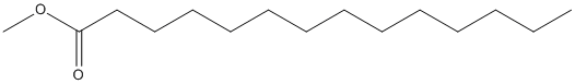
Esterases receive special attention because their wide distribution in biological systems and environments and their importance for physiology and chemical synthesis. The prediction of esterases substrate promiscuity level from sequence data and the molecular reasons why certain such enzymes are more promiscuous than others, remain to be elucidated. This limits the surveillance of the sequence space for esterases potentially leading to new versatile biocatalysts and new insights into their role in cellular function. Here we performed an extensive analysis of the substrate spectra of 145 phylogenetically and environmentally diverse microbial esterases, when tested with 96 diverse esters. We determined the primary factors shaping their substrate range by analyzing substrate range patterns in combination with structural analysis and protein-ligand simulations. We found a structural parameter that helps ranking (classifying) promiscuity level of esterases from sequence data at 94% accuracy. This parameter, the active site effective volume, exemplifies the topology of the catalytic environment by measuring the active site cavity volume corrected by the relative solvent accessible surface area (SASA) of the catalytic triad. Sequences encoding esterases with active site effective volumes (cavity volume/SASA) above a threshold show greater substrate spectra, which can be further extended in combination with phylogenetic data. This measure provides also a valuable tool for interrogating substrates capable of being converted. This measure, found to be transferred to phosphatases of the haloalkanoic acid dehalogenase superfamily and possibly other enzymatic systems, represents a powerful tool for low-cost bioprospecting for esterases with broad substrate ranges, in large scale sequence datasets.
        
Title: The Purification and Characterization of Lipases from Lasiodiplodia theobromae, and Their Immobilization and Use for Biodiesel Production from Coconut Oil Venkatesagowda B, Ponugupaty E, Barbosa-Dekker AM, Dekker RFH Ref: Appl Biochem Biotechnol, :, 2018 : PubMed
The coconut kernel-associated fungus, Lasiodiplodia theobromae VBE1, was grown on coconut cake with added coconut oil as lipase inducer under solid-state fermentation conditions. The extracellular-produced lipases were purified and resulted in two enzymes: lipase A (68,000 Da)-purified 25.41-fold, recovery of 47.1%-and lipase B (32,000 Da)-purified 18.47-fold, recovery of 8.2%. Both lipases showed optimal activity at pH 8.0 and 35 degrees C, were activated by Ca(2+), exhibited highest specificity towards coconut oil and p-nitrophenyl palmitate, and were stable in iso-octane and hexane. Ethanol supported higher lipase activity than methanol, and n-butanol inactivated both lipases. Crude lipase immobilized by entrapment within 4% (w/v) calcium alginate beads was more stable than the crude-free lipase preparation within the range pH 2.5-10.0 and 20-80 degrees C. The immobilized lipase preparation was used to catalyze the transesterification/methanolysis of coconut oil to biodiesel (fatty acyl methyl esters (FAMEs)) and was quantified by gas chromatography. The principal FAMEs were laurate (46.1%), myristate (22.3%), palmitate (9.9%), and oleate (7.2%), with minor amounts of caprylate, caprate, and stearate also present. The FAME profile was comparatively similar to NaOH-mediated transesterified biodiesel from coconut oil, but distinctly different to petroleum-derived diesel. This study concluded that Lasiodiplodia theobromae VBE1 lipases have potential for biodiesel production from coconut oil.
The production of biodiesel from a naturally isolated strain of Chlamydomonas was investigated. The microalgal strain was isolated from the rice paddy-field soil samples during a screening program. The identification was done using physiological and molecular approaches. After reaching the stationary phase of growth, the total content of the lipids was extracted. The extracted fatty acids were primarily esterified and then identified through TLC and GC/MS analysis. Several types of fatty acid methyl esters (FAMEs) were identified in the isolated microalga and the presence of at least nine FAMEs in Chlamydomonas sp. MCCS 026 was shown. The total fatty acid content of the isolated strain was 25%. The composition of fatty acids in the studied species of microalga was mainly docosanoic acid methyl ester, tetradecanoic acid methyl ester, hexadecanoic acid methyl ester and nonanoic acid methyl ester.