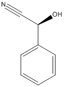
4 structures(e.g. : 6COE, 6COH, 6COI... more)(less)6COE: Structure of the hydroxynitrile lyase from Arabidopsis thaliana AtHNL enantioselectivity mutant At-A9-H7 Apo, Y13C,Y121L,P126F,L128W,C131T,F179L,A209I with benzaldehyde,mandelic acid nitrile, 6COH: Structure of the hydroxynitrile lyase from Arabidopsis thaliana AtHNL enantioselectivity mutant At-A9-H7 Apo, Y13C,Y121L,P126F,L128W,C131T,A209I with benzaldehyde, mandelic acid nitrile, 6COI: Structure of the hydroxynitrile lyase from Arabidopsis thaliana At-A9-H7 Apo, Y13C,Y121L,P126F,L128W,C131T,A209I with cyanide, benzaldehyde, mandelic acid nitrile, 1YB6: Hydroxynitrile lyase from hevea brasiliensis in complex with mandelonitrile
Title: Structural determinants of the enantioselectivity of the hydroxynitrile lyase from Hevea brasiliensis Gartler G, Kratky C, Gruber K Ref: J Biotechnol, 129:87, 2007 : PubMed
The hydroxynitrile lyase from the tropical rubber tree Hevea brasiliensis (HbHNL) is utilized as a biocatalyst in stereospecific syntheses of alpha-hydroxynitriles from aldehydes and methyl-ketones. The catalyzed reaction represents one of the few industrially relevant examples of enzyme mediated C-C coupling reactions. In this work, we determined the X-ray crystal structures (at 1.54 and 1.76 Angstroms resolution) of HbHNL complexes with two chiral substrates -- mandelonitrile and 2,3-dimethyl-2-hydroxy-butyronitrile -- by soaking and rapid freeze quenching techniques. This is the first structural observation of the complex between a HNL and chiral substrates. Consistent with the known selectivity of the enzyme, only the S-enantiomers of the two substrates were observed in the active site. The binding modes of the chiral substrates were identical to that observed for the biological substrate acetone cyanohydrin. This indicates that the transformation of these non-natural substrates follows the same mechanism. A large hydrophobic pocket was identified in the active site of HbHNL which accommodates the more voluminous substituents of the two substrates. A three-point binding mode of the substrates -- hydrophobic pocket, hydrogen bonds between the hydroxyl group and Ser80 and Thr11, electrostatic interaction of the cyano group with Lys236 -- offers a likely structural explanation for the enantioselectivity of the enzyme. The structural data rationalize the observed (S)-enantioselectivity and form the basis for modifying the stereospecificity through rational design. The structures also revealed the necessity of considerable flexibility of the sidechain of Trp128 in order to bind and transform larger substrates.
Tryptophan 128 of hydroxynitrile lyase of Manihot esculenta (MeHNL) covers a significant part of a hydrophobic channel that gives access to the active site of the enzyme. This residue was therefore substituted in the mutant MeHNL-W128A by alanine to study its importance for the substrate specificity of the enzyme. Wild-type MeHNL and MeHNL-W128A showed comparable activity on the natural substrate acetone cyanohydrin (53 and 40 U/mg, respectively). However, the specific activities of MeHNL-W128A for the unnatural substrates mandelonitrile and 4-hydroxymandelonitrile are increased 9-fold and approximately 450-fold, respectively, compared with the wild-type MeHNL. The crystal structure of the MeHNL-W128A substrate-free form at 2.1 A resolution indicates that the W128A substitution has significantly enlarged the active-site channel entrance, and thereby explains the observed changes in substrate specificity for bulky substrates. Surprisingly, the MeHNL-W128A--4-hydroxybenzaldehyde complex structure at 2.1 A resolution shows the presence of two hydroxybenzaldehyde molecules in a sandwich type arrangement in the active site with an additional hydrogen bridge to the reacting center.