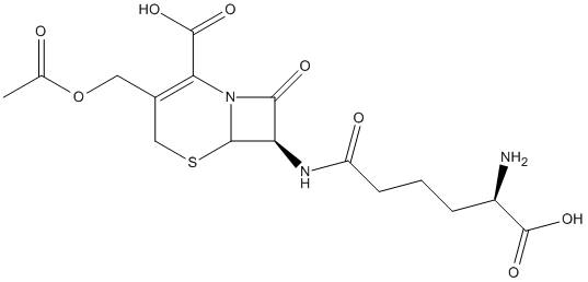
Organophosphorus insecticides and nerve agents irreversibly inhibit serine hydrolase superfamily enzymes. One enzyme of this superfamily, the industrially important (for beta-lactam antibiotic synthesis) AXE/CAH (acetyl xylan esterase/cephalosporin acetyl hydrolase) from the biotechnologically valuable organism Bacillus pumilus, exhibits low sensitivity to the organophosphate paraoxon (diethyl-p-nitrophenyl phosphate, also called paraoxon-ethyl), reflected in a high K(i) for it (~5 mM) and in a slow formation (t((1/2))~1 min) of the covalent adduct of the enzyme and for DEP (E-DEP, enzyme-diethyl phosphate, i.e. enzyme-paraoxon). The crystal structure of the E-DEP complex determined at 2.7 A resolution (1 A=0.1 nm) reveals strain in the active Ser(1)(8)(1)-bound organophosphate as a likely cause for the limited paraoxon sensitivity. The strain results from active-site-size limitation imposed by bulky conserved aromatic residues that may exclude as substrates esters having acyl groups larger than acetate. Interestingly, in the doughnut-like homohexamer of the enzyme, the six active sites are confined within a central chamber formed between two 60 degrees -staggered trimers. The exclusive access to this chamber through a hole around the three-fold axis possibly limits the size of the xylan natural substrates. The enzyme provides a rigid scaffold for catalysis, as reflected in the lack of movement associated with paraoxon adduct formation, as revealed by comparing this adduct structure with that also determined in the present study at 1.9 A resolution for the paraoxon-free enzyme.
        
Title: The last step in cephalosporin C formation revealed: crystal structures of deacetylcephalosporin C acetyltransferase from Acremonium chrysogenum in complexes with reaction intermediates Lejon S, Ellis J, Valegard K Ref: Journal of Molecular Biology, 377:935, 2008 : PubMed
Deacetylcephalosporin C acetyltransferase (DAC-AT) catalyses the last step in the biosynthesis of cephalosporin C, a broad-spectrum beta-lactam antibiotic of large clinical importance. The acetyl transfer step has been suggested to be limiting for cephalosporin C biosynthesis, but has so far escaped detailed structural analysis. We present here the crystal structures of DAC-AT in complexes with reaction intermediates, providing crystallographic snapshots of the reaction mechanism. The enzyme is found to belong to the alpha/beta hydrolase class of acetyltransferases, and the structures support previous observations of a double displacement mechanism for the acetyl transfer reaction in other members of this class of enzymes. The structures of DAC-AT reported here provide evidence of a stable acyl-enzyme complex, thus underpinning a mechanism involving acetylation of a catalytic serine residue by acetyl coenzyme A, followed by transfer of the acetyl group to deacetylcephalosporin C through a suggested tetrahedral transition state.
        
Title: Isolation, analysis, and expression of two genes from Thermoanaerobacterium sp. strain JW/SL YS485: a beta-xylosidase and a novel acetyl xylan esterase with cephalosporin C deacetylase activity Lorenz WW, Wiegel J Ref: Journal of Bacteriology, 179:5436, 1997 : PubMed
The genes encoding acetyl xylan esterase 1 (axe1) and a beta-xylosidase (xylB) have been cloned and sequenced from Thermoanaerobacterium sp. strain JW/SL YS485. axe1 is located 22 nucleotides 3' of the xylB sequence. The identity of axe1 was confirmed by comparison of the deduced amino acid sequence to peptide sequence analysis data from purified acetyl xylan esterase 1. The xylB gene was identified by expression cloning and by sequence homology to known beta-xylosidases. Plasmids which independently expressed either acetyl xylan esterase 1 (pAct1BK) or beta-xylosidase (pXylo-1.1) were constructed in Escherichia coli. Plasmid pXylAct-1 contained both genes joined at a unique EcoRI site and expressed both activities. Substrate specificity, pH, and temperature optima were determined for partially purified recombinant acetyl xylan esterase 1 and for crude recombinant beta-xylosidase. Similarity searches showed that the axe1 and xylB genes were homologs of the ORF-1 and xynB genes, respectively, isolated from Thermoanaerobacterium saccharolyticum. Although the deduced sequence of the axe1 product had no significant amino acid sequence similarity to any reported acetyl xylan esterase sequence, it did have strong similarity to cephalosporin C deacetylase from Bacillus subtilis. Recombinant acetyl xylan esterase 1 was found to have thermostable deacetylase activity towards a number of acetylated substrates, including cephalosporin C and 7-aminocephalosporanic acid.
Organophosphorus insecticides and nerve agents irreversibly inhibit serine hydrolase superfamily enzymes. One enzyme of this superfamily, the industrially important (for beta-lactam antibiotic synthesis) AXE/CAH (acetyl xylan esterase/cephalosporin acetyl hydrolase) from the biotechnologically valuable organism Bacillus pumilus, exhibits low sensitivity to the organophosphate paraoxon (diethyl-p-nitrophenyl phosphate, also called paraoxon-ethyl), reflected in a high K(i) for it (~5 mM) and in a slow formation (t((1/2))~1 min) of the covalent adduct of the enzyme and for DEP (E-DEP, enzyme-diethyl phosphate, i.e. enzyme-paraoxon). The crystal structure of the E-DEP complex determined at 2.7 A resolution (1 A=0.1 nm) reveals strain in the active Ser(1)(8)(1)-bound organophosphate as a likely cause for the limited paraoxon sensitivity. The strain results from active-site-size limitation imposed by bulky conserved aromatic residues that may exclude as substrates esters having acyl groups larger than acetate. Interestingly, in the doughnut-like homohexamer of the enzyme, the six active sites are confined within a central chamber formed between two 60 degrees -staggered trimers. The exclusive access to this chamber through a hole around the three-fold axis possibly limits the size of the xylan natural substrates. The enzyme provides a rigid scaffold for catalysis, as reflected in the lack of movement associated with paraoxon adduct formation, as revealed by comparing this adduct structure with that also determined in the present study at 1.9 A resolution for the paraoxon-free enzyme.
        
Title: The last step in cephalosporin C formation revealed: crystal structures of deacetylcephalosporin C acetyltransferase from Acremonium chrysogenum in complexes with reaction intermediates Lejon S, Ellis J, Valegard K Ref: Journal of Molecular Biology, 377:935, 2008 : PubMed
Deacetylcephalosporin C acetyltransferase (DAC-AT) catalyses the last step in the biosynthesis of cephalosporin C, a broad-spectrum beta-lactam antibiotic of large clinical importance. The acetyl transfer step has been suggested to be limiting for cephalosporin C biosynthesis, but has so far escaped detailed structural analysis. We present here the crystal structures of DAC-AT in complexes with reaction intermediates, providing crystallographic snapshots of the reaction mechanism. The enzyme is found to belong to the alpha/beta hydrolase class of acetyltransferases, and the structures support previous observations of a double displacement mechanism for the acetyl transfer reaction in other members of this class of enzymes. The structures of DAC-AT reported here provide evidence of a stable acyl-enzyme complex, thus underpinning a mechanism involving acetylation of a catalytic serine residue by acetyl coenzyme A, followed by transfer of the acetyl group to deacetylcephalosporin C through a suggested tetrahedral transition state.
A bromothymol blue-based colorimetric assay has been devised to screen for acetyl xylan esterase or cephalosporin C (CPC) deacetylase activities using 7-amino cephalosporanic acid (7-ACA), CPC, or acetylated xylan as substrate. These enzymes are not screened with their natural substrates because of the tedious procedures available previously. Acetyl xylan esterase from Bacillus pumilus CECT 5072 was cloned, expressed in Escherichia coli Rosetta (DE3), and characterized using this assay. Similar K(M) values for 7-ACA and CPC were obtained when compared with those described using HPLC methods. The assay is easy to perform and can be carried out in robotic high-throughput colorimetric devices normally used in directed evolution experiments. The assay allowed us to detect improvements in activity at a minimum of twofold with a very low coefficient of variance in 96-well plates. This method is significantly faster and more convenient to use than are known HPLC and pH-stat procedures.
        
Title: Batch production of deacetyl 7-aminocephalosporanic acid by immobilized cephalosporin-C deacetylase Takimoto A, Takakura T, Tani H, Yagi S, Mitsushima K Ref: Applied Microbiology & Biotechnology, 65:263, 2004 : PubMed
Bacillus subtilis SHS0133 cephalosporin-C deacetylase (CAH) overexpressed in Escherichia coli was immobilized on an anion-exchange resin, KA-890, using glutaraldehyde. The activity yield of immobilized enzyme was approximately 55% of the free enzyme. The pH range for stability of the immobilized enzyme (pH 5-10) was broader than that for free enzyme. The K(m)(app) value of immobilized enzyme for 7-aminocephalosporanic acid (7-ACA) was similar to that of the free enzyme. This immobilized enzyme obeyed Michaelis-Menten kinetics similar to those of the free enzyme. A batch-type reactor with a water jacket was employed for deacetylation of 7-ACA using CAH immobilized on KA-890. Ten kilograms of 7-ACA were completely converted to deacetyl 7-ACA at pH 8.0 within 90 min. The reaction kinetics agreed well with a computer simulation model. Moreover, the immobilized enzyme exhibited only a slight loss of the initial activity even after repeated use (52 times ) over a period of 70 days. This reaction will thus be useful for the production of cephalosporin-type antibiotics.
        
Title: High-level expression, purification, and some properties of a recombinant cephalosporin-C deacetylase Takimoto A, Yagi S, Mitsushima K Ref: J Biosci Bioeng, 87:456, 1999 : PubMed
To maximize the expression of the cephalosporin-C deacetylase (CAH) gene isolated from Bacillus subtilis SHS 0133 in Escherichia coli, a series of expression plasmids was constructed with various spacings between the Shine-Dalgarno sequence and the ATG initiation codon. As the most efficient expression plasmid, we selected pCAH431, which has the trp promoter, a replication origin derived from pAT153, and a spacing of 13 nucleotides. E. coli JM103 with pCAH431 produced 4.9 g of CAH per liter on cultivation at 37 degrees C for 20 h in a 30-l jar fermentor. Since the amount of CAH reached about 70% of the total protein in the soluble fraction of the cells, and CAH was recovered from the cell extracts in an active form, the CAH was purified easily to homogeneity by only one column chromatography step. Twenty grams of 7-aminocephalosporanic acid was completely converted to deacetyl-7-aminocephalosporanic acid, a starting material for cefcapene pivoxil hydrochloride, by 12 mg of the purified enzyme without significant appearance of by-products. Thus, our expression and purification system has made the industrial production of CAH possible.
        
Title: Isolation, analysis, and expression of two genes from Thermoanaerobacterium sp. strain JW/SL YS485: a beta-xylosidase and a novel acetyl xylan esterase with cephalosporin C deacetylase activity Lorenz WW, Wiegel J Ref: Journal of Bacteriology, 179:5436, 1997 : PubMed
The genes encoding acetyl xylan esterase 1 (axe1) and a beta-xylosidase (xylB) have been cloned and sequenced from Thermoanaerobacterium sp. strain JW/SL YS485. axe1 is located 22 nucleotides 3' of the xylB sequence. The identity of axe1 was confirmed by comparison of the deduced amino acid sequence to peptide sequence analysis data from purified acetyl xylan esterase 1. The xylB gene was identified by expression cloning and by sequence homology to known beta-xylosidases. Plasmids which independently expressed either acetyl xylan esterase 1 (pAct1BK) or beta-xylosidase (pXylo-1.1) were constructed in Escherichia coli. Plasmid pXylAct-1 contained both genes joined at a unique EcoRI site and expressed both activities. Substrate specificity, pH, and temperature optima were determined for partially purified recombinant acetyl xylan esterase 1 and for crude recombinant beta-xylosidase. Similarity searches showed that the axe1 and xylB genes were homologs of the ORF-1 and xynB genes, respectively, isolated from Thermoanaerobacterium saccharolyticum. Although the deduced sequence of the axe1 product had no significant amino acid sequence similarity to any reported acetyl xylan esterase sequence, it did have strong similarity to cephalosporin C deacetylase from Bacillus subtilis. Recombinant acetyl xylan esterase 1 was found to have thermostable deacetylase activity towards a number of acetylated substrates, including cephalosporin C and 7-aminocephalosporanic acid.