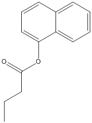
3 moreTitle: Identification and characterization of an acetyl xylan esterase from Aspergillus oryzae Kato T, Shiono Y, Koseki T Ref: J Biosci Bioeng, :, 2021 : PubMed
In this study, we report the identification and characterization of an acetyl xylan esterase, designated as AoAXEC, which was previously annotated as a hypothetical protein encoded by AO090023000158 in the Aspergillus oryzae genomic database. Based on its amino acid sequence, a low sequence identity to known acetyl xylan esterases was observed in the sequence of characterized acetyl xylan esterase. The gene fused with alpha-factor signal sequence of Saccharomyces cerevisiae instead of the native signal sequence was cloned into a vector, pPICZalphaC, and expressed successfully in Pichia pastoris as an active extracellular protein. The purified recombinant protein had pH and temperature optima of 7.0 and 50 degreesC, respectively, and was stable up to 50 degreesC. The optimal substrate for hydrolysis by the purified recombinant AoAXEC, among a panel of alpha-naphthyl esters (C2-C16), was alpha-naphthyl propionate (C3), with an activity of 0.35 +/- 0.006 units/mg protein. No significant difference of the K(m) value was observed between C3 (2.3 +/- 0.7 mM) and C2 (1.9 +/- 0.4 mM). In contrast, k(cat) value for C3 (18 +/- 3.9 s(-1)) was higher compared to C2 (4.5 +/- 0.7 s(-1)). The purified recombinant enzyme displayed a low activity toward acyl chain substrates containing eight or more carbon atoms. Recombinant AoAXEC catalyzed the release of acetic acid from wheat arabinoxylan. However, no activity was detected on methyl esters of ferulic, p-coumaric, caffeic, or sinapic acids. Additionally, the liberation of phenolic acids, such as ferulic acid, from wheat arabinoxylan was not exhibited by the recombinant protein.
Esterases receive special attention because their wide distribution in biological systems and environments and their importance for physiology and chemical synthesis. The prediction of esterases substrate promiscuity level from sequence data and the molecular reasons why certain such enzymes are more promiscuous than others, remain to be elucidated. This limits the surveillance of the sequence space for esterases potentially leading to new versatile biocatalysts and new insights into their role in cellular function. Here we performed an extensive analysis of the substrate spectra of 145 phylogenetically and environmentally diverse microbial esterases, when tested with 96 diverse esters. We determined the primary factors shaping their substrate range by analyzing substrate range patterns in combination with structural analysis and protein-ligand simulations. We found a structural parameter that helps ranking (classifying) promiscuity level of esterases from sequence data at 94% accuracy. This parameter, the active site effective volume, exemplifies the topology of the catalytic environment by measuring the active site cavity volume corrected by the relative solvent accessible surface area (SASA) of the catalytic triad. Sequences encoding esterases with active site effective volumes (cavity volume/SASA) above a threshold show greater substrate spectra, which can be further extended in combination with phylogenetic data. This measure provides also a valuable tool for interrogating substrates capable of being converted. This measure, found to be transferred to phosphatases of the haloalkanoic acid dehalogenase superfamily and possibly other enzymatic systems, represents a powerful tool for low-cost bioprospecting for esterases with broad substrate ranges, in large scale sequence datasets.
BACKGROUND: Microbial enzymes produced in the gastrointestinal tract are primarily responsible for the release and biochemical transformation of absorbable bioactive monophenols. In the present work we described the crystal structure of LJ0536, a serine cinnamoyl esterase produced by the probiotic bacterium Lactobacillus johnsonii N6.2. METHODOLOGY/PRINCIPAL FINDINGS: We crystallized LJ0536 in the apo form and in three substrate-bound complexes. The structure showed a canonical alpha/beta fold characteristic of esterases, and the enzyme is dimeric. Two classical serine esterase motifs (GlyXSerXGly) can be recognized from the amino acid sequence, and the structure revealed that the catalytic triad of the enzyme is formed by Ser(106), His(225), and Asp(197), while the other motif is non-functional. In all substrate-bound complexes, the aromatic acyl group of the ester compound was bound in the deepest part of the catalytic pocket. The binding pocket also contained an unoccupied area that could accommodate larger ligands. The structure revealed a prominent inserted alpha/beta subdomain of 54 amino acids, from which multiple contacts to the aromatic acyl groups of the substrates are made. Inserts of this size are seen in other esterases, but the secondary structure topology of this subdomain of LJ0536 is unique to this enzyme and its closest homolog (Est1E) in the Protein Databank. CONCLUSIONS: The binding mechanism characterized (involving the inserted alpha/beta subdomain) clearly differentiates LJ0536 from enzymes with similar activity of a fungal origin. The structural features herein described together with the activity profile of LJ0536 suggest that this enzyme should be clustered in a new group of bacterial cinnamoyl esterases.
        
3 lessTitle: Identification and characterization of an acetyl xylan esterase from Aspergillus oryzae Kato T, Shiono Y, Koseki T Ref: J Biosci Bioeng, :, 2021 : PubMed
In this study, we report the identification and characterization of an acetyl xylan esterase, designated as AoAXEC, which was previously annotated as a hypothetical protein encoded by AO090023000158 in the Aspergillus oryzae genomic database. Based on its amino acid sequence, a low sequence identity to known acetyl xylan esterases was observed in the sequence of characterized acetyl xylan esterase. The gene fused with alpha-factor signal sequence of Saccharomyces cerevisiae instead of the native signal sequence was cloned into a vector, pPICZalphaC, and expressed successfully in Pichia pastoris as an active extracellular protein. The purified recombinant protein had pH and temperature optima of 7.0 and 50 degreesC, respectively, and was stable up to 50 degreesC. The optimal substrate for hydrolysis by the purified recombinant AoAXEC, among a panel of alpha-naphthyl esters (C2-C16), was alpha-naphthyl propionate (C3), with an activity of 0.35 +/- 0.006 units/mg protein. No significant difference of the K(m) value was observed between C3 (2.3 +/- 0.7 mM) and C2 (1.9 +/- 0.4 mM). In contrast, k(cat) value for C3 (18 +/- 3.9 s(-1)) was higher compared to C2 (4.5 +/- 0.7 s(-1)). The purified recombinant enzyme displayed a low activity toward acyl chain substrates containing eight or more carbon atoms. Recombinant AoAXEC catalyzed the release of acetic acid from wheat arabinoxylan. However, no activity was detected on methyl esters of ferulic, p-coumaric, caffeic, or sinapic acids. Additionally, the liberation of phenolic acids, such as ferulic acid, from wheat arabinoxylan was not exhibited by the recombinant protein.
Esterases receive special attention because their wide distribution in biological systems and environments and their importance for physiology and chemical synthesis. The prediction of esterases substrate promiscuity level from sequence data and the molecular reasons why certain such enzymes are more promiscuous than others, remain to be elucidated. This limits the surveillance of the sequence space for esterases potentially leading to new versatile biocatalysts and new insights into their role in cellular function. Here we performed an extensive analysis of the substrate spectra of 145 phylogenetically and environmentally diverse microbial esterases, when tested with 96 diverse esters. We determined the primary factors shaping their substrate range by analyzing substrate range patterns in combination with structural analysis and protein-ligand simulations. We found a structural parameter that helps ranking (classifying) promiscuity level of esterases from sequence data at 94% accuracy. This parameter, the active site effective volume, exemplifies the topology of the catalytic environment by measuring the active site cavity volume corrected by the relative solvent accessible surface area (SASA) of the catalytic triad. Sequences encoding esterases with active site effective volumes (cavity volume/SASA) above a threshold show greater substrate spectra, which can be further extended in combination with phylogenetic data. This measure provides also a valuable tool for interrogating substrates capable of being converted. This measure, found to be transferred to phosphatases of the haloalkanoic acid dehalogenase superfamily and possibly other enzymatic systems, represents a powerful tool for low-cost bioprospecting for esterases with broad substrate ranges, in large scale sequence datasets.
In this study, we report the characterization of a protein from Aspergillus oryzae, exhibiting sequence identity with paraben esterase from the genus Aspergillus. The coding region of 1,586 bp, including a 77-bp intron, encoded a protein of 502 amino acids. The gene without the signal peptide of 19 amino acids was cloned into a vector, pPICZalphaC, and expressed successfully in Pichia pastoris as an active extracellular protein. The purified recombinant protein had pH and temperature optima of 7.0-8.0 and 30 degrees C, respectively, and was stable at the pH range of 7.0-10.0 and up to 40 degrees C. The optimal substrate for hydrolysis by the purified recombinant protein, among a panel of alpha-naphthyl esters (C2-C16), was alpha-naphthyl butyrate (C4), with activity of 0.16 units/mg protein. The considerable hydrolytic activity of the purified recombinant enzyme toward tributyrin was determined. However, no paraben esterase activity was detected toward the ethyl, propyl, and butyl esters of 4-hydroxybenzoic acid. In addition, no activity was detected toward the methyl esters of ferulic, p-coumaric, caffeic, and sinapic acids that would indicate feruloyl esterase activity.
BACKGROUND: Microbial enzymes produced in the gastrointestinal tract are primarily responsible for the release and biochemical transformation of absorbable bioactive monophenols. In the present work we described the crystal structure of LJ0536, a serine cinnamoyl esterase produced by the probiotic bacterium Lactobacillus johnsonii N6.2. METHODOLOGY/PRINCIPAL FINDINGS: We crystallized LJ0536 in the apo form and in three substrate-bound complexes. The structure showed a canonical alpha/beta fold characteristic of esterases, and the enzyme is dimeric. Two classical serine esterase motifs (GlyXSerXGly) can be recognized from the amino acid sequence, and the structure revealed that the catalytic triad of the enzyme is formed by Ser(106), His(225), and Asp(197), while the other motif is non-functional. In all substrate-bound complexes, the aromatic acyl group of the ester compound was bound in the deepest part of the catalytic pocket. The binding pocket also contained an unoccupied area that could accommodate larger ligands. The structure revealed a prominent inserted alpha/beta subdomain of 54 amino acids, from which multiple contacts to the aromatic acyl groups of the substrates are made. Inserts of this size are seen in other esterases, but the secondary structure topology of this subdomain of LJ0536 is unique to this enzyme and its closest homolog (Est1E) in the Protein Databank. CONCLUSIONS: The binding mechanism characterized (involving the inserted alpha/beta subdomain) clearly differentiates LJ0536 from enzymes with similar activity of a fungal origin. The structural features herein described together with the activity profile of LJ0536 suggest that this enzyme should be clustered in a new group of bacterial cinnamoyl esterases.
        
Title: Isolation, characterization, and heterologous expression of a carboxylesterase of Pseudomonas aeruginosa PAO1 Pesaresi A, Devescovi G, Lamba D, Venturi V, Degrassi G Ref: Curr Microbiol, 50:102, 2005 : PubMed
We purified to homogeneity an intracellular esterase from the opportunistic pathogen Pseudomonas aeruginosa PAO1. The enzyme hydrolyzes p-nitrophenyl acetate and other acetylated substrates. The N-terminal amino acid sequence was analyzed and 11 residues, SEPLILDAPNA, were determined. The corresponding gene PA3859 was identified in the P. aeruginosa PAO1 genome as the only gene encoding for a protein with this N-terminus. The encoding gene was cloned in Escherichia coli, and the recombinant protein expressed and purified to homogeneity. According to sodium dodecyl sulfate-polyacrylamide gel electrophoresis (SDS-PAGE) analysis and analytical gel filtration chromatography, the esterase was found to be a monomer of approximately 24 kDa. The experimentally determined isoelectric point was 5.2 and the optimal enzyme activity was at 55 degrees C and at pH 9.0. The esterase preferentially hydrolyzed short-chain fatty acids. It is inhibited by phenylmethylsulfonyl fluoride (PMSF) but not by ethylendiaminotetraacetic acid (EDTA). Native enzyme preparations typically showed a Michaelis constant (K(m)) and V(max) of 0.43 mM and 12,500 U mg(-1), respectively, using p-nitrophenyl acetate as substrate. Homology-based database searches clearly revealed the presence of the consensus GXSXG signature motif that is present in the serine-dependent acylhydrolase protein family.
        
Title: Cloning and analysis of CUT1, a cutinase gene from Magnaporthe grisea Sweigard JA, Chumley FG, Valent B Ref: Molecular & General Genetics, 232:174, 1992 : PubMed
A gene from Magnaporthe grisea was cloned using a cDNA clone of the Colletotrichum gloeosporioides cutinase gene as a heterologous probe; the nucleotide sequence of a 2 kb DNA segment containing the gene has been determined. DNA hybridization analysis shows that the M. grisea genome contains only one copy of this gene. The predicted polypeptide contains 228 amino acids and is homologous to the three previously characterized cutinases, showing 74% amino acid similarity to the cutinase of C. gloeosporioides. Comparison with previously determined cutinase sequences suggests that the gene contains two introns, 115 and 147 bp in length. The gene is expressed when cutin is the sole carbon source but not when the carbon source is cutin and glucose together or glucose alone. Levels of intracellular and extracellular cutinase activity increase in response to growth in the presence of cutin. The activity level is higher in a transformant containing multiple copies of the cloned gene than in the parent strain. Non-denaturing polyacrylamide gels stained for esterase activity show a single major band among intracellular and extracellular proteins from cutin-grown cultures that is not present among intracellular and extracellular proteins prepared from glucose-grown or carbon-starved cultures. This band stains more intensely in extracts from the multicopy transformant than in extracts from the parent strain. We conclude that the cloned DNA contains a M. grisea gene for cutinase, which we have named CUT1.