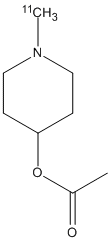
As a neuromodulator, the neurotransmitter acetylcholine plays an important role in cognitive, mood, locomotor, sleep/wake, and olfactory functions. In the pathophysiology of most neurodegenerative diseases, such as Alzheimer disease (AD) or Lewy body disorder (LBD), cholinergic receptors, transporters, or enzymes are involved and relevant as imaging targets. The aim of this review is to summarize current knowledge on PET imaging of cholinergic neurotransmission in neurodegenerative diseases. For PET imaging of presynaptic vesicular acetylcholine transporters (VAChT), (-)-(18)F-fluoroethoxybenzovesamicol ((18)F-FEOBV) was the first PET ligand that could be successfully translated to clinical application. Since then, the number of (18)F-FEOBV PET investigations on patients with AD or LBD has grown rapidly and provided novel, important findings concerning the pathophysiology of AD and LBD. Regarding the alpha4beta2 nicotinic acetylcholine receptors (nAChRs), various second-generation PET ligands, such as (18)F-nifene, (18)F-AZAN, (18)F-XTRA, (-)-(18)F-flubatine, and (+)-(18)F-flubatine, were developed and successfully translated to human application. In neurodegenerative diseases such as AD and LBD, PET imaging of alpha4beta2 nAChRs is of special value for monitoring disease progression and drugs directed to alpha4beta2 nAChRs. For PET of alpha7 nAChR, (18)F-ASEM and (11)C-MeQAA were successfully applied in mild cognitive impairment and AD, respectively. The highest potential for alpha7 nAChR PET is seen in staging, in evaluating disease progression, and in therapy monitoring. PET of selective muscarinic acetylcholine receptors (mAChRs) is still in an early stage, as the development of subtype-selective radioligands is complicated. Promising radioligands to image mAChR subtypes M1 ((11)C-LSN3172176), M2 ((18)F-FP-TZTP), and M4 ((11)C-MK-6884) were developed and successfully translated to humans. PET imaging of mAChRs is relevant for the assessment and monitoring of therapies in AD and LBD. PET of acetylcholine esterase activity has been investigated since the 1990s. Many PET studies with (11)C-PMP and (11)C-MP4A demonstrated cortical cholinergic dysfunction in dementia associated with AD and LBD. Recent studies indicated a solid relationship between subcortical and cortical cholinergic dysfunction and noncognitive dysfunctions such as balance and gait in LBD. Taken together, PET of distinct components of cholinergic neurotransmission is of great interest for diagnosis, disease monitoring, and therapy monitoring and to gain insight into the pathophysiology of different neurodegenerative disorders.
        
Title: Use of a novel radiometric method to assess the inhibitory effect of donepezil on acetylcholinesterase activity in minimally diluted tissue samples Kikuchi T, Okamura T, Arai T, Obata T, Fukushi K, Irie T, Shiraishi T Ref: British Journal of Pharmacology, 159:1732, 2010 : PubMed
BACKGROUND AND PURPOSE: Cholinesterase inhibitors have been widely used for the treatment of patients with dementia. Monitoring of the cholinesterase activity in the blood is used as an indicator of the effect of the cholinesterase inhibitors in the brain. The selective measurement of cholinesterase with low tissue dilution is preferred for accurate monitoring; however, the methods have not been established. Here, we investigated the effect of tissue dilution on the action of cholinesterase inhibitors using a novel radiometric method with selective substrates, N-[(14)C]methylpiperidin-4-yl acetate ([(14)C]MP4A) and (R)-N- [(14)C]methylpiperidin-3-yl butyrate ([(14)C]MP3B_R), for AChE and butyrylcholinesterase (BChE) respectively. EXPERIMENTAL APPROACH: We investigated the kinetics of hydrolysis of [(14)C]-MP4A and [(14)C]-MP3B_R by cholinesterases, and evaluated the selectivity of [(14)C]MP4A and [(14)C]MP3B_R for human AChE and BChE, respectively, compared with traditional substrates. Then, IC(50) values of cholinesterase inhibitors in minimally diluted and highly diluted tissues were measured with [(14)C]MP4A and [(14)C]MP3B_R. KEY RESULTS: AChE and BChE activities were selectively measured as the first-order hydrolysis rates of [(14)C]-MP4A and [(14)C]MP3B_R respectively. The AChE selectivity of [(14)C]MP4A was an order of magnitude higher than traditional substrates used for the AChE assay. The IC(50) values of specific AChE and BChE inhibitors, donepezil and ethopropazine, in 1.2-fold diluted human whole blood were much higher than those in 120-fold diluted blood. In addition, the IC(50) values of donepezil in monkey brain were dramatically decreased as the tissue was diluted. CONCLUSIONS AND IMPLICATIONS: This method would effectively monitor the activity of cholinesterase inhibitors used for therapeutics, pesticides and chemical warfare agents.
        
Title: [Imaging of brain acetylcholinesterase activity in dementias and extrapyramidal disorders] Shinotoh H Ref: Rinsho Shinkeigaku, 47:822, 2007 : PubMed
Carbon-11 labeled N-methylpiperidin-4-yl acetate ([11C]MP4A) and carbon-11 labeled N-methylpiperidin-4-yl propionate ([11C]MP4P) are acetylcholine analogues and have been successfully used for measurement of brain acetylcholinesterase (AChE) activity in vivo in humans. In Alzheimer's disease (AD), there is a significant loss of AChE activity in the cerebral cortex in association with mental decline. The reduction of AChE activity in the cerebral cortex is more remarkable in early-onset AD than late-onset AD. There is mild but significant reduction of AChE activity in the cerebral cortex, even in the early stage of Parkinson's disease (PD) without dementia. There is remarkable reduction of AChE activity in the entire cerebral cortex in both PD with dementia and dementia with Lewy bodies (DLB). In two patients with frontotemporal dementia with parkinsonism linked to chromosome 17, there was prominent reduction of AChE activity in the cerebral cortex and thalamus. In 12 patients with progressive supranuclear palsy, there was profound reduction of AChE activity in the thalamus but not in the cerebral cortex. In corticobasal degeneration, there is symmetrical loss of AChE activity in the sensori-motor cortex.
As a neuromodulator, the neurotransmitter acetylcholine plays an important role in cognitive, mood, locomotor, sleep/wake, and olfactory functions. In the pathophysiology of most neurodegenerative diseases, such as Alzheimer disease (AD) or Lewy body disorder (LBD), cholinergic receptors, transporters, or enzymes are involved and relevant as imaging targets. The aim of this review is to summarize current knowledge on PET imaging of cholinergic neurotransmission in neurodegenerative diseases. For PET imaging of presynaptic vesicular acetylcholine transporters (VAChT), (-)-(18)F-fluoroethoxybenzovesamicol ((18)F-FEOBV) was the first PET ligand that could be successfully translated to clinical application. Since then, the number of (18)F-FEOBV PET investigations on patients with AD or LBD has grown rapidly and provided novel, important findings concerning the pathophysiology of AD and LBD. Regarding the alpha4beta2 nicotinic acetylcholine receptors (nAChRs), various second-generation PET ligands, such as (18)F-nifene, (18)F-AZAN, (18)F-XTRA, (-)-(18)F-flubatine, and (+)-(18)F-flubatine, were developed and successfully translated to human application. In neurodegenerative diseases such as AD and LBD, PET imaging of alpha4beta2 nAChRs is of special value for monitoring disease progression and drugs directed to alpha4beta2 nAChRs. For PET of alpha7 nAChR, (18)F-ASEM and (11)C-MeQAA were successfully applied in mild cognitive impairment and AD, respectively. The highest potential for alpha7 nAChR PET is seen in staging, in evaluating disease progression, and in therapy monitoring. PET of selective muscarinic acetylcholine receptors (mAChRs) is still in an early stage, as the development of subtype-selective radioligands is complicated. Promising radioligands to image mAChR subtypes M1 ((11)C-LSN3172176), M2 ((18)F-FP-TZTP), and M4 ((11)C-MK-6884) were developed and successfully translated to humans. PET imaging of mAChRs is relevant for the assessment and monitoring of therapies in AD and LBD. PET of acetylcholine esterase activity has been investigated since the 1990s. Many PET studies with (11)C-PMP and (11)C-MP4A demonstrated cortical cholinergic dysfunction in dementia associated with AD and LBD. Recent studies indicated a solid relationship between subcortical and cortical cholinergic dysfunction and noncognitive dysfunctions such as balance and gait in LBD. Taken together, PET of distinct components of cholinergic neurotransmission is of great interest for diagnosis, disease monitoring, and therapy monitoring and to gain insight into the pathophysiology of different neurodegenerative disorders.
BACKGROUND: Alzheimer's disease (AD) is a neurodegenerative disorder characterized by chronic progressive cognitive decline and displays underlying brain cholinergic dysfunction, providing a rationale for treatment with cholinomimetic medication. The clinical presentations and courses of AD patients may differ by age of onset. OBJECTIVE: The objective of the present study was to illustrate the regional differences of brain acetylcholinesterase (AChE) activity as quantified by N-[11C]methylpiperidinyl-4-acetate ([11C]MP4A) and PET using parametric whole brain analysis and clarify those differences as a function of age. METHODS: 22 early onset AD (EOAD) with age at onset under 65, the remaining 26 as late onset AD (LOAD), and 16 healthy controls (HC) were enrolled. Voxel-based AChE activity estimation of [11C]MP4A PET images was conducted by arterial input and unconstrained nonlinear least-squares method with subsequent parametrical analyses. Statistical threshold was set as Family Wise Error corrected, p-value <0.05 on cluster-level and cluster extent over 30 voxels. RESULTS: Voxel-based group comparison showed that, compared to HC, both EOAD and LOAD showed cortical AChE decrement in parietal, temporal, and occipital cortices, with wider and stringent cortical involvement in the EOAD group, most prominently demonstrated in the temporal region. There was no significant correlation between age and regional cerebral AChE activity except for a small left superior temporal region in the AD group (Brodmann's area 22, Zmax = 5.13, 396 voxels), whereas no significant cluster was found in the HC counterpart. CONCLUSION: Difference in cortical cholinergic dysfunction between EOAD and LOAD may shed some light on the cholinomimetic drug efficacy in AD.
        
Title: Dementia with Lewy bodies can be well-differentiated from Alzheimer's disease by measurement of brain acetylcholinesterase activity-[(11)C]MP4A PET study Bohnen NI, Muller ML, Kaufer DI Ref: Int J Geriatr Psychiatry, 31:951, 2016 : PubMed
Evidence for cholinergic dysfunction in very early stages of neurodegeneration like mild cognitive impairment (MCI) is inconclusive. Previous positron emission tomography (PET) studies based on small samples investigated if it is related to memory impairment. We examined whether cortical acetylcholine esterase (AChE) activity is reduced at this stage and correlated with cognitive function. N-[(11)C]-methyl-4-piperidyl acetate ([11C]MP4A), a positron emission tomography tracer for measuring cerebral AChE activity in vivo, was applied in 21 controls and 17 MCI patients. Parametric images of AChE activity were analyzed using standard atlas regions. Principal components analysis (PCA) of regional values of AChE activity and correlation analysis with neuropsychological test results was performed. Cortical AChE activity showed a significant decline in MCI patients compared with controls which was most pronounced in temporal regions. They formed the main part of a principal component that was related significantly to verbal and nonverbal memory, language comprehension and executive function. Cholinergic dysfunction is an early hallmark even before onset of dementia at the clinical stage of MCI. Its impact especially on temporal neocortex is associated with impaired neuropsychological function.
CONTEXT: Both neuropsychological and functional magnetic resonance imaging studies have shown deficiencies in face perception in subjects with autism spectrum disorders (ASD). The fusiform gyrus has been regarded as the key structure in face perception. The cholinergic system is known to regulate the function of the visual pathway, including the fusiform gyrus. OBJECTIVES: To determine whether central acetylcholinesterase activity, a marker for the cholinergic system, is altered in ASD and whether the alteration in acetylcholinesterase activity, if any, is correlated with their social functioning. DESIGN: Using positron emission tomography and a radiotracer, N-[(11)C]methyl-4-piperidyl acetate ([(11)C]MP4A), regional cerebrocortical acetylcholinesterase activities were estimated by reference tissue-based linear least-squares analysis and expressed in terms of the rate constant k(3). Current and childhood autism symptoms in the adult subjects with ASD were assessed by the Autism Diagnostic Observation Schedule and the Autism Diagnostic Interview-Revised, respectively. Voxel-based analyses as well as region of interest-based methods were used for between-subject analysis and within-subject correlation analysis with respect to clinical variables. SETTING: Participants recruited from the community. PARTICIPANTS: Twenty adult subjects with ASD (14 male and 6 female; age range, 18-33 years; mean [SD] intelligence quotient, 91.6 [4.3]) and 20 age-, sex-, and intelligence quotient-matched healthy controls. RESULTS: Both voxel- and region of interest-based analyses revealed significantly lower [(11)C]MP4A k(3) values in the bilateral fusiform gyri of subjects with ASD than in those of controls (P < .05, corrected). The fusiform k(3) values in subjects with ASD were negatively correlated with their social disabilities as assessed by Autism Diagnostic Observation Schedule as well as Autism Diagnostic Interview-Revised. CONCLUSIONS: The results suggest that a deficit in cholinergic innervations of the fusiform gyrus, which can be observed in adults with ASD, may be related to not only current but also childhood impairment of social functioning.
        
Title: Use of a novel radiometric method to assess the inhibitory effect of donepezil on acetylcholinesterase activity in minimally diluted tissue samples Kikuchi T, Okamura T, Arai T, Obata T, Fukushi K, Irie T, Shiraishi T Ref: British Journal of Pharmacology, 159:1732, 2010 : PubMed
BACKGROUND AND PURPOSE: Cholinesterase inhibitors have been widely used for the treatment of patients with dementia. Monitoring of the cholinesterase activity in the blood is used as an indicator of the effect of the cholinesterase inhibitors in the brain. The selective measurement of cholinesterase with low tissue dilution is preferred for accurate monitoring; however, the methods have not been established. Here, we investigated the effect of tissue dilution on the action of cholinesterase inhibitors using a novel radiometric method with selective substrates, N-[(14)C]methylpiperidin-4-yl acetate ([(14)C]MP4A) and (R)-N- [(14)C]methylpiperidin-3-yl butyrate ([(14)C]MP3B_R), for AChE and butyrylcholinesterase (BChE) respectively. EXPERIMENTAL APPROACH: We investigated the kinetics of hydrolysis of [(14)C]-MP4A and [(14)C]-MP3B_R by cholinesterases, and evaluated the selectivity of [(14)C]MP4A and [(14)C]MP3B_R for human AChE and BChE, respectively, compared with traditional substrates. Then, IC(50) values of cholinesterase inhibitors in minimally diluted and highly diluted tissues were measured with [(14)C]MP4A and [(14)C]MP3B_R. KEY RESULTS: AChE and BChE activities were selectively measured as the first-order hydrolysis rates of [(14)C]-MP4A and [(14)C]MP3B_R respectively. The AChE selectivity of [(14)C]MP4A was an order of magnitude higher than traditional substrates used for the AChE assay. The IC(50) values of specific AChE and BChE inhibitors, donepezil and ethopropazine, in 1.2-fold diluted human whole blood were much higher than those in 120-fold diluted blood. In addition, the IC(50) values of donepezil in monkey brain were dramatically decreased as the tissue was diluted. CONCLUSIONS AND IMPLICATIONS: This method would effectively monitor the activity of cholinesterase inhibitors used for therapeutics, pesticides and chemical warfare agents.
        
Title: Development of Alzheimer's disease imaging agents for clinical studies Ryu EK, Chen X Ref: Front Biosci, 13:777, 2008 : PubMed
Alzheimer's disease (AD) is a neurodegenerative disease characterized by a progressive loss of neurotransmitters, as well as acetylcholinesterase and nicotinic acetylcholine receptors in the central nervous system that leads to learning and memory deficits, among other problems. The disease is associated with increased production and accumulation extracellular amyloid plaques and neurofibrillary tangles in aging human brain, shown in postmortem exams. New methods for reliable in vivo measurement of brain therefore would be much more ideal. PET and SPECT imaging are sensitive methods for the quantitation of AD biomarkers. The development of molecular imaging agents for AD is critically important in the early diagnosis, neuropathogenesis studies and treatment of AD. A number of potential diagnostic PET and SPECT imaging agents targeting AD have been synthesized and evaluated. Although many agents showed excellent results for in vitro monitoring of the disease, there are only several radioligands with high selectivity and specificity to binding sites and appropriate pharmacokinetics, such as [11C]MP4A, [11C]PMP, [11C]nicotine, 2- or 6-[18F]fluoro-A-85380, [11C]SB-13, [11C]PIB, and [18F]FDDNP, that have been tested in AD patients. Here we review some recent progress and development of AD imaging agents using PET and SPECT in human clinical studies.
Cerebral acetylcholinesterase (AChE) imaging is not only useful for diagnosis of dementia disorders but also for therapeutic monitoring of the effects of cholinesterase (ChE) inhibitors and for decision of the appropriate clinical dosage of newly developed ChE inhibitors. Several ChE inhibitors or the derivatives such as 1,2,3,4-tetrahydro-9-methylaminoacridine (MTHA), donepezil, physostigmine, CP126,998 and 2-fluoro-CP118,954 have been labeled with positron emitters for mapping cerebral AChE by positron emission tomography (PET). When [(11)C]MTHA or [(11)C]donepezil was injected in animals, the uptake poorly reflect the regional distribution of AChE in the brain because of high non-specific binding and/or less specific to AChE in vivo in the brain tissue. [(11)C]physostigmine, [(11)C]CP126,998 and 2-[(18)F]fluoro-CP118,954 were distributed corresponding well to the regional AChE activity in animals, and also former two probes were successfully applied to clinical PET trial. The other approach is measuring cerebral AChE activity with radiolabeled acetylcholine analogue substrates. We have developed the principle for measuring cerebral enzyme activity by PET and radiolabeled N-methylpiperidinyl esters for quantitative measurement of cerebral AChE activity. N-[(11)C]methylipiperidin-4-yl acetate (MP4A) and N-[(11)C]methylpiperidin-4-yl propionate (MP4P) have been used for clinical studies of other demented disorders including Alzheimer's disease (AD), and the probes have demonstrated not only the reduction of AChE activity in the cerebral cortex of patients with AD but also the inhibitory effects of donepezil and rivastigmine on AChE activity in the brain of AD patients. Following this succession, widely available [(18)F]-labeled derivatives of MP4A and MP4P have been developed based on the structure-activity relationships between AChE and piperidinol esters.
        
Title: [Imaging of brain acetylcholinesterase activity in dementias and extrapyramidal disorders] Shinotoh H Ref: Rinsho Shinkeigaku, 47:822, 2007 : PubMed
Carbon-11 labeled N-methylpiperidin-4-yl acetate ([11C]MP4A) and carbon-11 labeled N-methylpiperidin-4-yl propionate ([11C]MP4P) are acetylcholine analogues and have been successfully used for measurement of brain acetylcholinesterase (AChE) activity in vivo in humans. In Alzheimer's disease (AD), there is a significant loss of AChE activity in the cerebral cortex in association with mental decline. The reduction of AChE activity in the cerebral cortex is more remarkable in early-onset AD than late-onset AD. There is mild but significant reduction of AChE activity in the cerebral cortex, even in the early stage of Parkinson's disease (PD) without dementia. There is remarkable reduction of AChE activity in the entire cerebral cortex in both PD with dementia and dementia with Lewy bodies (DLB). In two patients with frontotemporal dementia with parkinsonism linked to chromosome 17, there was prominent reduction of AChE activity in the cerebral cortex and thalamus. In 12 patients with progressive supranuclear palsy, there was profound reduction of AChE activity in the thalamus but not in the cerebral cortex. In corticobasal degeneration, there is symmetrical loss of AChE activity in the sensori-motor cortex.
Donepezil hydrochloride is a potent and selective inhibitor for brain acetylcholinesterase (AChE) and is currently used worldwide for the treatment of Alzheimer's disease. Until now, there is no in vivo study on the relation between the plasma concentration and the brain AChE inhibition. The purpose of this study was to estimate in vivo plasma IC(50) of donepezil in living monkeys by measuring plasma donepezil concentration (LC/MS/MS) and brain AChE activity with positron emission tomography (PET) and N-[(11)C]methylpiperidin-4-yl acetate, which is an acetylcholine analog recently developed by us for quantifying in vivo brain AChE activity. PET scans with donepezil at two doses, 100 microg/kg (donepezil-1; N=5) or 250 microg/kg (donepezil-2; N=5), were performed using the same monkeys at 4-week intervals. Before each PET scan, baseline PET scans (N=10 in total) were performed without donepezil. The plasma donepezil concentrations 14 min after intravenous injection were proportional to the doses, 17.2+/-2.9 ng/ml (donepezil-1) and 44.0+/-5.0 ng/ml (donepezil-2), and the mean AChE inhibitions in four neocortical regions as evaluated by PET were also dose-dependent, 27% (donepezil-1) and 53% (donepezil-2). In IC(50) estimation, measured plasma donepezil concentrations were corrected for the change during PET scan. The IC(50) values (estimate+/-SE) were 42+/-9.0 (ng/ml; donepezil-1), 34+/-3.2 (donepezil-2), and 37+/-4.1 (combined data). The present method may be useful for in vivo evaluation of other AChE inhibitors and novel drugs.
        
Title: Acetylcholinesterase imaging: its use in therapy evaluation and drug design Shinotoh H, Fukushi K, Nagatsuka S, Irie T Ref: Curr Pharm Des, 10:1505, 2004 : PubMed
Several cholinesterase (ChE) inhibitors have been labeled with carbon-11 for visualizing binding sites on acetylcholinesterase (AChE) by positron emission tomography (PET). Following intravenous injection of 1,2,3,4-tetrahydro-9-[(11)C]methylaminoacridine or [(11)C]donepezil, however, the radioactivity distribution does not reflect the regional distribution of AChE in the brain of animals, probably because these compounds have high non-specific binding and/or other specific binding sites in vivo in the brain. PET studies with [(11)C]physostigmine and [(11)C]CP-126,998 in the brain of healthy subjects have shown a radioactivity distribution corresponding to the regional distribution of AChE activity measured in postmortem human brains. These radiotracers may be useful for measuring the occupancy of binding sites on AChE by AChE inhibitors, and for investigating the cerebral pharmacokinetics of such therapeutic drugs. An alternative approach to map AChE is the use of acetylcholine analogue substrates. We have developed N-methylpiperidinyl esters labeled with carbon-11 for quantitative measurement of AChE activity. Currently, two N-[(11)C]methylpiperidine esters, N-[(11)C]methylipiperidin-4-ylacetate (MP4A) and N-[(11)C]methylpiperidin-4-yl propionate (MP4P or PMP), have been used for clinical studies of Alzheimer's disease and other neurodegenerative diseases. Both [(11)C]MP4A- and [(11)C]MP4P-PET have demonstrated not only the reduction of AChE activity in the cerebral cortex of patients with Alzheimer's disease (AD) but also the inhibitory effects of donepezil and rivastigmine on AChE activity in the brain of AD patients. AChE imaging should prove useful for therapeutic monitoring of the effects of ChE inhibitors, including determination of the appropriate clinical doses of newly developed compounds, and can thus prompt the development of novel drugs targeting AChE.
        
Title: Effects of acute acetylcholinesterase inhibition on the cerebral cholinergic neuronal system and cognitive function: Functional imaging of the conscious monkey brain using animal PET in combination with microdialysis Tsukada H, Nishiyama S, Fukumoto D, Ohba H, Sato K, Kakiuchi T Ref: Synapse, 52:1, 2004 : PubMed
This study demonstrated the effects of acute acetylcholinesterase (AChE) inhibition by donepezil (Aricept) on the cerebral cholinergic neuronal system in the brains of young (5.2 +/- 1.1 years old) and aged (20.3 +/- 2.6 years old) monkeys (Macaca mulatta) in the conscious state. Donepezil at doses of 50 and 250 microg/kg suppressed AChE activity, analyzed by metabolic rate (k(3)) of N-[(11)C]methyl-4-piperidyl acetate ([(11)C]MP4A), in all cortical regions in a dose-dependent manner in both age groups. However, the suppression degree was more marked in young than in aged monkeys. AChE inhibition by donepezil resulted in a dose-dependent increase in acetylcholine levels in the prefrontal cortex of young animals as measured by microdialysis. Binding of (+)N-[(11)C]propyl-3-piperidyl benzilate ([(11)C](+)3-PPB) to cortical muscarinic receptors was reduced by donepezil, probably in a competitive inhibition manner. Aged monkeys showed less reduction of [(11)C](+)3-PPB binding than young animals. As evaluated by an oculomotor delayed response task, aged monkeys showed impaired working memory performance compared to young monkeys, and the impaired performance was partly improved by the administration of donepezil, due to the facilitation of the cholinergic neuronal system by AChE inhibition. These results demonstrate that the PET imaging technique with specific labeled compounds in combination with microdialysis and a behavioral cognition task could be a useful method to clarify the mechanism of drugs in the living brains of experimental animals.
OBJECTIVE Brain acetylcholinesterase activity was determined in healthy controls and in patients with mild cognitive impairment and early Alzheimer's disease.
METHODS:
A specific acetylcholinesterase tracer, [methyl-(11)C]N-methyl-piperidyl-4-acetate ([(11)C]MP4A), and a three dimensional PET system with magnetic resonance coregistration were used for imaging.
RESULTS:
There was a significant difference in the acetylcholinesterase activity in the hippocampus between the groups (p = 0.03), the mean (SD) acetylcholinesterase activity (k(3) values, min(-1)) being 0.114 (0.036) in controls, 0.098 (0.023) in mild cognitive impairment, and 0.085 (0.022) in Alzheimer's disease. The mini-mental state examination score showed no significant relation with acetylcholinesterase activity in any brain area in the combined mild cognitive impairment/Alzheimer group.
CONCLUSIONS:
Hippocampal acetylcholinesterase activity is only slightly reduced in mild cognitive impairment and early Alzheimer's disease and so the value of in vivo acetylcholinesterase measurements in detecting the early Alzheimer process is limited.
A new method for quantitative measurement of brain acetylcholinesterase (AChE) activity in living human brain using positron emission tomography (PET) is described. We tested several radiolabeled lipophilic acetylcholine analogs, e.g., N-methylpiperidyl esters, which readily entered the brain via the blood-brain barrier, were hydrolyzed selectively by AChE, and were then trapped in the brain. Among them, and tested and N-[11C]methylpiperidin-4-yl acetate ([11C]MP4A) was chosen as the tracer for PET. Quantitative measurement of cortical AChE was accomplished by fitting the time course of cerebral radioactivity concentration measured by PET and the metabolite-corrected arterial plasma input function using a nonlinear least-squares fitting method. Normal control studies of subjects with a wide range in age (24-89 years) showed no decrease in AChE activity in the cerebral cortex with age. Studies on patients with Alzheimer's disease demonstrated a widespread reduction of AChE activity in the cerebral cortex (more profound in early-onset than in late-onset Alzheimer's disease). Parkinson's disease and progressive supranuclear palsy, clinically similar disorders, could be differentiated with [11C]MP4A/PET studies. Simple methods without using an arterial input function are also proposed. The method provides a quantitative measure of the cholinergic aspect of brain function and proved to be useful in diagnosis of neurodegenerative disorders including Alzheimer's disease.
N-[18F]Fluoroethyl-4-piperidyl acetate ([18F]FEtP4A), an analog of [11C]MP4A for mapping brain acetylcholineseterase (AchE) activity, was prepared by reacting 4-piperidyl acetate (P4A) with [18F]fluoroethyl bromide ([18F]FEtBr) using a newly developed automated system. Preliminary evaluation showed that the initial uptake of [18F]FEtP4A in the mouse brain was > 8% injected dose/g tissue. The distribution pattern of [18F]FEtP4A in the brain was striatum>cerebral cortex>cerebellum within 10-120 min post-injection, which reflected the distribution rank pattern of AchE activity in the brain. Moreover, chemical analysis of in vivo radioactive metabolites in the mouse brain indicated that 83% of [18F]FEtP4A was hydrolyzed to N-[18F]fluoroethyl-4-piperidinol ([18F]FEtP4OH) after 1 min intravenous injection. From these results, [18F]FEtP4A may become a promising PET tracer for mapping the AchE in vivo.
        
Title: Kinetic analysis of [(11)C]MP4A using a high-radioactivity brain region that represents an integrated input function for measurement of cerebral acetylcholinesterase activity without arterial blood sampling Nagatsuka Si S, Fukushi K, Shinotoh H, Namba H, Iyo M, Tanaka N, Aotsuka A, Ota T, Tanada S, Irie T Ref: Journal of Cerebral Blood Flow & Metabolism, 21:1354, 2001 : PubMed
N -[(11)C]methylpiperidin-4-yl acetate ([(11)C]MP4A) is an acetylcholine analog. It has been used successfully for the quantitative measurement of acetylcholinesterase (AChE) activity in the human brain with positron emission tomography (PET). [(11)C]MP4A is specifically hydrolyzed by AChE in the brain to a hydrophilic metabolite, which is irreversibly trapped locally in the brain. The authors propose a new method of kinetic analysis of brain AChE activity by PET without arterial blood sampling, that is, reference tissue-based linear least squares (RLS) analysis. In this method, cerebellum or striatum is used as a reference tissue. These regions, because of their high AChE activity, act as a biologic integrator of plasma input function during PET scanning, when regional metabolic rates of [(11)C]MP4A through AChE (k(3); an AChE index) are calculated by using Blomqvist's linear least squares analysis. Computer simulation studies showed that RLS analysis yielded k(3) with almost the same accuracy as the standard nonlinear least squares (NLS) analysis in brain regions with low (such as neocortex and hippocampus) and moderately high (thalamus) k(3) values. The authors then applied these methods to [(11) C]MP4A PET data in 12 healthy subjects and 26 patients with Alzheimer disease (AD) using the cerebellum as the reference region. There was a highly significant linear correlation in regional k(3) estimates between RLS and NLS analyses (456 cerebral regions, [RLS k(3) ] = 0.98 x [NLS k(3) ], r = 0.92, P < 0.001). Significant reductions were observed in k(3) estimates of frontal, temporal, parietal, occipital, and sensorimotor cerebral neocortices (P < 0.001, single-tailed t-test), and hippocampus (P = 0.012) in patients with AD as compared with controls when using RLS analysis. Mean reductions (19.6%) in these 6 regions by RLS were almost the same as those by NLS analysis (20.5%). The sensitivity of RLS analysis for detecting cortical regions with abnormally low k 3 in the 26 patients with AD (138 of 312 regions, 44%) was somewhat less than NLS analysis (52%), but was greater than shape analysis (33%), another method of [(11)C]MP4A kinetic analysis without blood sampling. The authors conclude that RLS analysis is practical and useful for routine analysis of clinical [(11)C]MP4A studies.
        
Title: Positron emission tomographic measurement of brain acetylcholinesterase activity using N-[(11)C]methylpiperidin-4-yl acetate without arterial blood sampling: methodology of shape analysis and its diagnostic power for Alzheimer's disease Tanaka N, Fukushi K, Shinotoh H, Nagatsuka S, Namba H, Iyo M, Aotsuka A, Ota T, Tanada S, Irie T Ref: Journal of Cerebral Blood Flow & Metabolism, 21:295, 2001 : PubMed
N-[11C]methylpiperidin-4-yl acetate ([11C]MP4A) is a radiotracer that has been used successfully for the quantitative measurement of acetylcholinesterase (AChE) activity in the human brain with positron emission tomography (PET) using a standard compartment model analysis and a metabolite-corrected arterial input function. In the current study, the authors evaluated the applicability of a simple kinetic analysis without blood sampling, namely shape analysis. First, the authors used computer simulations to analyze factors that affect the precision and bias of shape analysis, then optimized the shape analysis procedure for [11C]MP4A. Before shape analysis execution, the later part of dynamic PET data except for the initial 3 minutes were smoothed by fitting to a bi-exponential function followed by linear interpolation of 8 data points between each of adjacent scan frames. Simulations showed that shape analysis yielded estimates of regional metabolic rates of [11C]MP4A by AChE (k3) with acceptable precision and bias in brain regions with low k3 values such as neocortex. Estimates in regions with higher k3 values became progressively more inaccurate. The authors then applied the method to [11C]MP4A PET data in 10 healthy subjects and 20 patients with Alzheimer's disease (AD). There was a highly significant linear correlation in regional k3 estimates between shape and compartment analyses (300 neocortical regions, [shape k3] = 0.93 x [NLS k3], r = 0.89, P < 0.001). Significant reductions in k3 estimates of frontal, temporal, parietal, occipital, and sensorimotor cerebral cortices in patients with AD as compared with controls were observed when using shape analysis (P < 0.013, two-tailed t-test), although these reductions (17% to 20%) were somewhat less than those obtained by compartment analysis (22% to 27%). The sensitivity of shape analysis for detecting neocortical regions with abnormally low k3 in the 20 patients with AD (92 out of 200 regions, 46%) also was somewhat less than compartment analysis (136 out of 200 regions, 68%). However, taking its simplicity and noninvasiveness into account, the authors conclude that quantitative measurement of neocortical AChE activity with shape analysis and [11C]MP4A PET is practical and useful for clinical diagnosis of AD.
The regional cerebral metabolic rate of [11C]N-methyl-4-piperidyl acetate, which is nearly proportional to regional cerebral acetylcholinesterase (AChE) activity, was measured by dynamic positron emission tomography in 20 healthy subjects with a wide age range (24-89 years). Quantitative measurement was achieved using a kinetic model which consisted of arterial plasma and cerebral tissue compartments. The plasma input function was obtained using thin-layer chromatography and an imaging phosphor plate system at frequent sampling intervals to catch the rapid metabolism of the tracer in the blood. The distribution of the rate constant k3, an index of AChE activity, agreed well with reported post-mortem AChE distribution in the cerebral cortex (0.067-0.097 min-1) and thalamus (0.268 min-1), where AChE activity was low to moderate. The k3 values in the striatum and cerebellum, where AChE activity was very high, did not respond linearly to AChE activity because of increased flow dependency. No significant effect of age was found on AChE activity of the cerebral cortex, suggesting that the ascending central cholinergic system is preserved in normal aging. This study has shown that quantitative measurement of enzyme activity in the living brain is possible through appropriate modelling of tracer kinetics and accurate measurement of the input function. The method should be applicable to patients with Alzheimer's disease and those with other kinds of dementia whose central cholinergic system has been reported to be disturbed.
We measured brain acetylcholinesterase activity in 16 patients with Parkinson's disease (PD), 12 patients with progressive supranuclear palsy (PSP), and 13 age-matched controls, using N-methyl-4-[11C]piperidyl acetate and positron emission tomography. Kinetic analysis was performed to calculate k3, an index of acetylcholinesterase activity. In PD patients, there was a significant reduction (-17%) of cerebral cortical k3 compared with normal controls, whereas there was only a nonsignificant reduction (-10%) of cortical k3 in PSP patients. However, there was a prominent reduction (-38%) of thalamic k3 in PSP patients compared with normal controls, whereas there was only a nonsignificant reduction (-13%) of thalamic k3 in PD patients. The results suggest that there is a loss of cholinergic innervation to the cerebral cortex in association with cholinergic innervation to the thalamus in PD, whereas there is a preferential loss of cholinergic innervation to the thalamus in PSP. When the thalamic to cerebral cortical k3 ratio was taken for each subject, PD and PSP were separated, suggesting that positron emission tomography measurement of acetylcholinesterase activity may be useful for differentiating the two similar disorders.
        
Title: N-[11C]methylpiperidine esters as acetylcholinesterase substrates: an in vivo structure-reactivity study Kilbourn MR, Nguyen TB, Snyder SE, Sherman P Ref: Nucl Med Biol, 25:755, 1998 : PubMed
A series of simple esters incorporating the N-[11C]methylpiperidine structure were examined as in vivo substrates for acetylcholinesterase in mouse brain. 4-N-[11C]Methylpiperidinyl esters, including the acetate, propionate and isobutyrate esters, are good in vivo substrates for mammalian cholinesterases. Introduction of a methyl group at the 4-position of the 4-piperidinol esters, to form the ester of a teritary alcohol, effectively blocks enzymatic action. Methylation of 4-N-[11C]methylpiperidinyl propionate at the 3-position gives a derivative with increased in vivo reactivity toward acetylcholinesterase. Esters of piperidinecarboxylic acids (nipecotic, isonipecotic and pipecolinic acid ethyl esters) are not hydrolyzed by acetylcholinesterase in vivo, nor do they act as in vivo inhibitors of the enzyme. This study has identified simple methods to both increase and decrease the in vivo reactivity of piperidinyl esters toward acetylcholinesterase.
        
Title: Syntheses of carbon-11 labeled piperidine esters as potential in vivo substrates for acetylcholinesterase Nguyen TB, Snyder SE, Kilbourn MR Ref: Nucl Med Biol, 25:761, 1998 : PubMed
A series of carbon-11 labeled N-methylpiperidinyl esters were prepared as potential in vivo substrates for acetylcholinesterase (AChE). Target compounds were designed based on the structure of N-[11C]methylpiperidin-4-yl propionate, an ester currently used to measure AChE enzymatic activity in the human brain, to examine the structure-activity relationship for in vivo enzymatic hydrolysis. Changes in steric bulk and in the ester order ("reverse" esters) were made. Addition of methyl groups was made to both the acid side chain (synthesis of N-[11C]methylmethylpiperidin-4-yl isobutyrate) and to the piperidine ring (syntheses of N-[11C]methyl-4-methylpiperidin-4-yl propionate, N-[11C]methyl-4-methylpiperidin-4-yl acetate, and N-[11C]methyl-3-methylpiperidin-4-yl propionate). Alterations of the order of the ester heteroatoms was accomplished through syntheses of the N-[11C]methyl-2,3- and 4-piperidinecarboxylic acid ethyl esters. Finally, an additional piperidine-based ester (N-[11C]methylpiperidin-2-yl)methyl propionate was also prepared. All carbon-11-labeled esters were prepared by N-[11C]methylation reactions, using the desmethyl precursors and no-carrier-added [11C]methyltriflate, and were obtained in decay-corrected yields (not optimized) of 10-40% and high specific activities.
BACKGROUND:
Acetylcholinesterase activity, a marker for degeneration of the central cholinergic system, has consistently been reported, in necropsy brain studies, to be reduced in the cerebral cortex of patients with Alzheimer's disease. We have shown regional acetylcholinesterase activity in vivo in rodent and primate brains with radioactive acetylcholine analogues. In the present study, we used one of the analogues to map acetylcholinesterase activity in the brains of living people.
METHODS:
Positron emission tomography (PET) and a radiolabelled acetylcholine analogue with high hydrolytic specificity to acetylcholinesterase [11C]N-methyl-4-piperidyl acetate (MP4A), was used in eight elderly healthy controls and five patients with Alzheimer's disease who had mild dementia. All participants were given an intravenous injection of [11C]MP4A and then sequential patterns of radioactivity in various brain regions were obtained by PET. Time courses of [11C]MP4A concentration in arterial blood were also measured to obtain an input function. A three-compartment model was used to estimate regional acetylcholinesterase activity in the brain.
FINDINGS:
The estimated acetylcholinesterase distribution in the brain of the control participants agreed with the acetylcholinesterase distribution at necropsy. All patients with Alzheimer's disease had multiple cortical regions with a reduced estimated acetylcholinesterase activity in comparison with control participants. The reduction was more pronounced in the parietotemporal cortex, with an average reduction rate of 31% in temporal and 38% in parietal cortex, and less pronounced in other cortical lesions (19% in frontal, 24% in occipital, and 20% in sensorimotor cortex). Each patient was found to have at least two cortical regions with significantly reduced acetylcholinesterase activity.
INTERPRETATION:
The method we describe for non-invasive in-vivo detection of regional acetylcholinesterase changes in the living human brain that is feasible for biochemical assessment of Alzheimer's disease.