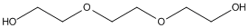
Structure-based drug design utilizes apo-protein or complex structures retrieved from the PDB. >57% of crystallographic PDB entries were obtained with polyethyleneglycols (PEGs) as precipitant and/or as cryoprotectant, but >6% of these report presence of individual ethyleneglycol oligomers. We report a case in which ethyleneglycol oligomers' presence in a crystal structure markedly affected the bound ligand's position. Specifically, we compared the positions of methylene blue and decamethonium in acetylcholinesterase complexes obtained using isomorphous crystals precipitated with PEG200 or ammonium sulfate. The ligands' positions within the active-site gorge in complexes obtained using PEG200 are influenced by presence of ethyleneglycol oligomers in both cases bound to W84 at the gorge's bottom, preventing interaction of the ligand's proximal quaternary group with its indole. Consequently, both ligands are approximately 3.0A further up the gorge than in complexes obtained using crystals precipitated with ammonium sulfate, in which the quaternary groups make direct pi-cation interactions with the indole. These findings have implications for structure-based drug design, since data for ligand-protein complexes with polyethyleneglycol as precipitant may not reflect the ligand's position in its absence, and could result in selecting incorrect drug discovery leads. Docking methylene blue into the structure obtained with PEG200, but omitting the ethyleneglycols, yields results agreeing poorly with the crystal structure; excellent agreement is obtained if they are included. Many proteins display features in which precipitants might lodge. It will be important to investigate presence of precipitants in published crystal structures, and whether it has resulted in misinterpreting electron density maps, adversely affecting drug design. This article is protected by copyright. All rights reserved.
        
Title: Structural and mutational analyses of Aes, an inhibitor of MalT in Escherichia coli Schiefner A, Gerber K, Brosig A, Boos W Ref: Proteins, 82:268, 2014 : PubMed
The acyl esterase Aes effectively inhibits the transcriptional activity of MalT-the central activator of maltose and maltodextrin utilizing genes in Escherichia coli. To provide better insight into the nature of the interaction between Aes and MalT, we determined two different crystal structures of Aes-in its native form and covalently modified by a phenylmethylsulfonyl moiety at its active site serine. Both structures show distinct space groups and were refined to a resolution of 1.8 A and 2.3 A, respectively. The overall structure of Aes resembles a canonical alpha/beta-hydrolase fold, which is extended by a funnel-like cap structure that forms the substrate-binding site. The catalytic triad of Aes, comprising residues Ser165, His292, and Asp262, is located at the bottom of this funnel. Analysis of the crystal-packing contacts of the two different space groups as well as analytical size-exclusion chromatography revealed a homodimeric arrangement of Aes. The Aes dimer adopts an antiparallel contact involving both the hydrolase core and the cap, with its twofold axis perpendicular to the largest dimension of Aes. To identify the surface area of Aes that is responsible for the interaction with MalT, we performed a structure-based alanine-scanning mutagenesis to pinpoint Aes residues that are significantly impaired in MalT inhibition, but still exhibit wild-type expression and enzymatic activity. These residues map to a shallow slightly concave surface patch of Aes at the opposite site of the dimerization interface and indicate the surface area that interacts with MalT. Proteins 2014; 82:268-277. (c) 2013 Wiley Periodicals, Inc.