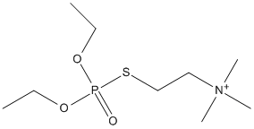
A potent, long-acting irreversible cholinesterase inhibitor used as an ocular hypertensive in the treatment of glaucoma. Occasionally used for accomodative esotropia.
Enzyme-catalyzed hydrolysis of echothiophate, a P-S bonded organophosphorus (OP) model, was spectrofluorimetrically monitored, using Calbiochem Probe IV as the thiol reagent. OP hydrolases were: the G117H mutant of human butyrylcholinesterase capable of hydrolyzing OPs, and a multiple mutant of Brevundimonas diminuta phosphotriesterase, GG1, designed to hydrolyze a large spectrum of OPs at high rate, including V agents. Molecular modeling of interaction between Probe IV and OP hydrolases (G117H butyrylcholinesterase, GG1, wild types of Brevundimonas diminuta and Sulfolobus solfataricus phosphotriesterases, and human paraoxonase-1) was performed. The high sensitivity of the method allowed steady-state kinetic analysis of echothiophate hydrolysis by highly purified G117H butyrylcholinesterase concentration as low as 0.85 nM. Hydrolysis was michaelian with Km = 0.20 +/- 0.03 mM and kcat = 5.4 +/- 1.6 min(-1). The GG1 phosphotriesterase hydrolyzed echothiophate with a high efficiency (Km = 2.6 +/- 0.2 mM; kcat = 53400 min(-1)). With a kcat/Km = (2.6 +/- 1.6) x 10(7) M(-1)min(-1), GG1 fulfills the required condition of potential catalytic bioscavengers. quantum mechanics/molecular mechanics (QM/MM) and molecular docking indicate that Probe IV does not interact significantly with the selected phosphotriesterases. Moreover, results on G117H mutant show that Probe IV does not inhibit butyrylcholinesterase. Therefore, Probe IV can be recommended for monitoring hydrolysis of P-S bonded OPs by thiol-free OP hydrolases.
        
Title: Site-directed mutagenesis of active site residues reveals plasticity of human butyrylcholinesterase in substrate and inhibitor interactions Gnatt A, Loewenstein Y, Yaron A, Schwarz M, Soreq H Ref: Journal of Neurochemistry, 62:749, 1994 : PubMed
In search of the molecular mechanisms underlying the broad substrate and inhibitor specificities of butyrylcholinesterase (BCHE), we employed site-directed mutagenesis to modify the catalytic triad residue Ser198, the acyl pocket Leu286 and adjacent Phe329 residues, and Met437 and Tyr440 located near the choline binding site. Mutant proteins were produced in microinjected Xenopus oocytes, and Km values towards butyrylthiocholine and IC50 values for the organophosphates diisopropylfluorophosphonate (DFP), diethoxyphosphinylthiocholine iodide (echothiophate), and tetraisopropylpyrophosphoramide (iso-OMPA) were determined. Substitution of Ser198 by cysteine and Met437 by aspartate nearly abolished activity, and other mutations of Ser198 completely abolished it. Tyr440 and Leu286 mutants remained active, but with higher Km and IC50 values. Rates of inhibition by DFP were roughly parallel to IC50 values for several Leu286 mutants. Both Km and IC50 values increased for Leu286 mutants in the order Asp < Gln < Lys. In contrast, cysteine, leucine, and glutamine mutants of Phe329 displayed unmodified Km values toward butyrylthiocholine, but up to 10-fold decreased IC50 values for DFP, iso-OMPA, and echothiophate. These findings add Tyr440 and Phe329 to the list of residues interacting with substrate and ligands, demonstrate plasticity in the active site region of BCHE, and foreshadow the design of recombinant BCHEs with tailored scavenging properties.
Human pancreas contains two cholinesterase isoenzymes: acetylcholinesterase (AChE) and butyrylcholinesterase (BCHE). In the present study, binding potency of two organophosphates for human cholinesterases were compared by the Ellman method. Echothiophate was found to have much greater potency than iso-OMPA for both cholinesterases. Using Karnovsky histochemical stains on human pancreatic tissue, the same results were confirmed. Dose-response studies with acetylcholine were done on viable pancreas fragments from nine human donors, without pancreatic disease (group I). Cold-preservation time was less than 30 h. Pancreas was minced into fragments, after the technique of Scheele and Palade, placed in Eagle's medium, and gassed with O2. Amylase release was measured by the Phadebas Method and corrected for basal release. There was a dose-dependent response to acetylcholine at 1 and 2 h, with a shift in peak amylase release to the left, when fragments were preincubated in 10(-4) M echothiophate. This indicated a 100-fold increase in sensitivity to acetylcholine. In three patients with chronic pancreatitis (Group II), there were variable patterns of response of amylase release to acetylcholine, and higher basal outputs. In Group III, prolonged storage conditions of over 40 h were tested for 4 pancreas donor tissues. There was no response to acetylcholine. These studies show that for up to 30 h cold storage, fragments of pancreas from human organ donors respond to acetylcholine in dose-dependent manner. An organophosphate, echothiophate (10(-4) M) which inhibits both cholinesterases, increases pancreatic sensitivity to acetylcholine, and these results are similar to findings from canine pancreas fragments, which also showed increased sensitivity.
Enzyme-catalyzed hydrolysis of echothiophate, a P-S bonded organophosphorus (OP) model, was spectrofluorimetrically monitored, using Calbiochem Probe IV as the thiol reagent. OP hydrolases were: the G117H mutant of human butyrylcholinesterase capable of hydrolyzing OPs, and a multiple mutant of Brevundimonas diminuta phosphotriesterase, GG1, designed to hydrolyze a large spectrum of OPs at high rate, including V agents. Molecular modeling of interaction between Probe IV and OP hydrolases (G117H butyrylcholinesterase, GG1, wild types of Brevundimonas diminuta and Sulfolobus solfataricus phosphotriesterases, and human paraoxonase-1) was performed. The high sensitivity of the method allowed steady-state kinetic analysis of echothiophate hydrolysis by highly purified G117H butyrylcholinesterase concentration as low as 0.85 nM. Hydrolysis was michaelian with Km = 0.20 +/- 0.03 mM and kcat = 5.4 +/- 1.6 min(-1). The GG1 phosphotriesterase hydrolyzed echothiophate with a high efficiency (Km = 2.6 +/- 0.2 mM; kcat = 53400 min(-1)). With a kcat/Km = (2.6 +/- 1.6) x 10(7) M(-1)min(-1), GG1 fulfills the required condition of potential catalytic bioscavengers. quantum mechanics/molecular mechanics (QM/MM) and molecular docking indicate that Probe IV does not interact significantly with the selected phosphotriesterases. Moreover, results on G117H mutant show that Probe IV does not inhibit butyrylcholinesterase. Therefore, Probe IV can be recommended for monitoring hydrolysis of P-S bonded OPs by thiol-free OP hydrolases.
        
Title: Effects of organophosphates on cholinesterase activity and neurite regeneration in Aplysia Srivatsan M Ref: Chemico-Biological Interactions, 119-120:371, 1999 : PubMed
In Aplysia, a marine mollusc, acetylcholinesterase (AChE) is present in cholinergic and non-cholinergic neurons and in hemolymph. Aplysia hemolymph has a very high level of AChE which promotes neurite growth in primary cultures of dopaminergic neurons via a non-catalytic mechanism. In contrast, AChE is known to facilitate neurite growth in cholinoceptive neurons by hydrolyzing ACh which inhibits neurite growth. In order to test whether AChE's site-specific neurotrophic action varies with the neuronal phenotype, we investigated the effects of active-site inhibited hemolymph AChE on neurite growth of cholinergic neurons of Aplysia in primary culture. Organophosphates being long-acting active site inhibitors of AChE were chosen for this study. The effects of active site inhibited hemolymph AChE was tested on large cholinergic neurons, R2 (abdominal ganglion) and LPL1 (left pleural ganglion) as well as small cholinergic neurons (buccal ganglion) of Aplysia, maintained in culture. Partially purified hemolymph AChE was inhibited by either 10 microM of echothiophate or 5 microM of paraoxon. Neurons were maintained in (1) L15 (defined medium) alone; (2) L15 + echothiophate; (3) L-15 + paraoxon; (4) L-15 + hemolymph AChE; (5) L15 + hemolymph AChE + echothiophate; and (6) L-15 + hemolymph AChE + paraoxon. Addition of uninhibited hemolymph AChE significantly increased neurite growth of cultured neurons compared to L15 alone. In the presence of echothiophate-inhibited or praoxon-inhibited AChE, neurite growth was significantly reduced when compared to L15 + uninhibited AChE. While the presence of echothiophate by itself did not reduce survival or neurite growth when compared to L-15 alone, the presence of paraoxon by itself markedly reduced survival and neurite growth of cultured neurons. The results show that AChE's catalytic action contributes to enhance neurite growth in cholinergic neurons and the effects of paraoxon appears to differ from that of echothiophate on cholinergic neurons of Aplysia.
        
Title: A comparison of the electrophysiological effects of two organophosphates, mipafox and ecothiopate, on mouse limb muscles de Blaquiere GE, Williams FM, Blain PG, Kelly SS Ref: Toxicol Appl Pharmacol, 150:350, 1998 : PubMed
Adult male albino mice were given single subcutaneous injections of either mipafox (110 mumol/kg) or ecothiopate (0.5 mumol/kg), two organophosphorus compounds (OPs). Acetylcholinesterase activity was measured in the soleus (slow-twitch) and extensor digitorum longus (EDL; fast-twitch) muscles. At 7 and 28 days after dosing, in vitro electrophysiological measurements were carried out in the soleus and EDL. Action potentials and end-plate potentials were evoked at 30 Hz and recorded intracellularly from single muscle fibers. The amplitudes, time course, and latencies of these potentials were measured and the variability (jitter) of latencies was calculated. Recordings after mipafox were also made with 3-Hz stimulation. Acetylcholinesterase activity was inhibited by mipafox (65% in the soleus; 76% in the EDL) and ecothiopate (59% in the soleus; 42% in the EDL). Mipafox and ecothiopate both increased postjunctional (muscle action potential) jitter in the soleus and EDL at 7 days after dosing. Organophosphates caused an increase in end-plate potential amplitudes in the soleus. Mipafox caused an increase in prejunctional (end-plate potential) jitter at 28 days after dosing in both muscles. A single dose of ecothiopate also caused an increase in prejunctional jitter at 28 days in the soleus. The OP-induced increase in jitter was different at different frequencies of stimulation. The results show that there are electrophysiological changes in both muscles after administration of organophosphorus compounds. The slow-twitch soleus appears more sensitive to prejunctional changes caused by OPs than the fast-twitch EDL.
        
Title: Importance of aspartate-70 in organophosphate inhibition, oxime re-activation and aging of human butyrylcholinesterase Masson P, Froment MT, Bartels CF, Lockridge O Ref: Biochemical Journal, 325:53, 1997 : PubMed
Asp-70 is the defining amino acid in the peripheral anionic site of human butyrylcholinesterase (BCHE), whereas acetylcholinesterase has several additional amino acids, the most important one being Trp-277 (Trp-279 in Torpedo AChE). We studied mutants D70G, D70K and A277W to evaluate the role of Asp-70 and Trp-277 in reactions with organophosphates. We found that Asp-70 was important for binding positively charged echothiophate, but not neutral paraoxon and iso-OMPA. Asp-70 was also important for binding of positively charged pralidoxime (2-PAM) and for activation of re-activation by excess 2-PAM. Excess 2-PAM had an effect similar to substrate activation, suggesting the binding of 2 mol of 2-PAM to wild-type but not to the D70G mutant. A surprising result was that Asp-70 was important for irreversible aging, the D70G mutant having a 3- and 8-fold lower rate of aging for paraoxon-inhibited and di-isopropyl fluorophosphate-inhibited BCHE. Mutants of Asp-70 had the same rate constants for phosphorylation and re-activation by 2-PAM as wild-type. The A277W mutant behaved like wild-type in all assays. Our results predict that people with the atypical (D70G) variant of BCHE will be more sensitive to the toxic effects of echothiophate, but will be equally sensitive to paraoxon and di-isopropyl fluorophosphate. People with the D70G mutation will be resistant to re-activation of their inhibited BCHE by 2-PAM, but this will be offset by the lower rate of irreversible aging of inhibited BCHE, allowing some regeneration by spontaneous hydrolysis.
        
Title: Acetylcholinesterase promotes regeneration of neurites in cultured adult neurons of Aplysia Srivatsan M, Peretz B Ref: Neuroscience, 77:921, 1997 : PubMed
Aplysia, a marine mollusc, has significant amounts of acetylcholinesterase in its hemolymph, reaching maximum levels in the adults with reproductive maturity [Srivatsan M., et al. (1992) J. comp. Physiol. 162, 29-37]. Since hemolymph of mature Aplysia is neurotrophic to Aplysia neurons in culture [Schacher S. and Proshanski E. (1983) J. Neurosci. 3, 2403-2413], we examined whether acetylcholinesterase is a hemolymph neurotrophic factor. Dopaminergic neurons from the pedal ganglia of young adult Aplysia were maintained in culture in defined medium or defined medium supplemented with hemolymph. After 24 h, neurons in defined medium supplemented with hemolymph were well attached to the substratum and exhibited multiple, long neurites. In contrast, neurons in defined medium alone attached poorly and exhibited one or two short neurites. When acetylcholinesterase was inhibited with a specific, membrane-impermeable inhibitor (1,5-bis(4-allyldimethylammoniumphenyl)-pentan-3-one dibromide) which binds to its catalytic and peripheral anionic sites, the neurotrophic effect of hemolymph was significantly reduced. However, inhibition of the catalytic site alone with membrane impermeable echothiophate still resulted in enhanced neurite growth. An analogue of acetylcholine, carbachol, which is not hydrolysed by acetylcholinesterase, did not interfere with neurite growth when added to the supplemented medium. Acetylcholinesterase isolated from the hemolymph and highly purified human acetylcholinesterase also promoted neurite growth in Aplysia neurons. These results show that i) acetylcholinesterase circulating in the hemolymph promotes neurite growth of adult neurons in culture; ii) the growth promoting action of acetylcholinesterase is independent of its function of hydrolysing acetylcholine and iii) the peripheral anionic site of acetylcholinesterase appears to be involved in neurite regeneration.
        
Title: Cholinergic toxicity resulting from ocular instillation of echothiophate iodide eye drops Manoguerra A, Whitney C, Clark RF, Anderson B, Turchen S Ref: Journal of Toxicology Clinical Toxicology, 33:463, 1995 : PubMed
A patient developed a severe cholinergic syndrome from the use of echothiophate iodide ophthalmic drops, presented with profound muscle weakness and was initially given the diagnosis of myasthenia gravis. Red blood cell and serum cholinesterase levels were severely depressed and symptoms resolved spontaneously following discontinuation of the eye drops.
        
Title: Site-directed mutagenesis of active site residues reveals plasticity of human butyrylcholinesterase in substrate and inhibitor interactions Gnatt A, Loewenstein Y, Yaron A, Schwarz M, Soreq H Ref: Journal of Neurochemistry, 62:749, 1994 : PubMed
In search of the molecular mechanisms underlying the broad substrate and inhibitor specificities of butyrylcholinesterase (BCHE), we employed site-directed mutagenesis to modify the catalytic triad residue Ser198, the acyl pocket Leu286 and adjacent Phe329 residues, and Met437 and Tyr440 located near the choline binding site. Mutant proteins were produced in microinjected Xenopus oocytes, and Km values towards butyrylthiocholine and IC50 values for the organophosphates diisopropylfluorophosphonate (DFP), diethoxyphosphinylthiocholine iodide (echothiophate), and tetraisopropylpyrophosphoramide (iso-OMPA) were determined. Substitution of Ser198 by cysteine and Met437 by aspartate nearly abolished activity, and other mutations of Ser198 completely abolished it. Tyr440 and Leu286 mutants remained active, but with higher Km and IC50 values. Rates of inhibition by DFP were roughly parallel to IC50 values for several Leu286 mutants. Both Km and IC50 values increased for Leu286 mutants in the order Asp < Gln < Lys. In contrast, cysteine, leucine, and glutamine mutants of Phe329 displayed unmodified Km values toward butyrylthiocholine, but up to 10-fold decreased IC50 values for DFP, iso-OMPA, and echothiophate. These findings add Tyr440 and Phe329 to the list of residues interacting with substrate and ligands, demonstrate plasticity in the active site region of BCHE, and foreshadow the design of recombinant BCHEs with tailored scavenging properties.
        
Title: Correlation of the anticholinesterase activity of a series of organophosphates with their ability to compete with agonist binding to muscarinic receptors Ward TR, Ferris DJ, Tilson HA, Mundy WR Ref: Toxicology & Applied Pharmacology, 122:300, 1993 : PubMed
Some compounds that inhibit acetylcholinesterase (AChE) activity compete directly with quinuclidinyl benzilate (QNB) binding, a muscarinic antagonist which binds to all subtypes equally, and with cis-methyldioxolane (CD), an agonist that binds with high affinity to the M2 subtype of muscarinic receptors. The relationship between inhibition of AChE activity and the capability to affect muscarinic receptors directly has not been systematically explored. The interaction of eight organophosphates with muscarinic receptors was compared to their ability to inhibit AChE activity in vitro in tissue homogenates from rat hippocampus and frontal cortex, two cholinergically enriched areas of the brain. Of the compounds tested only echothiophate competed for [3H]QNB binding and only at concentrations greater than 100 microM. The anticholinesterase compounds were also tested for their ability to compete with a muscarinic receptor agonist, [3H]CD, which binds with high affinity (approximate KD = 3.5 nM) to 10 and 3% of the muscarinic receptors in the frontal cortex and hippocampus, respectively. The anticholinesterase compounds inhibited high-affinity [3H]CD binding up to 80% and the effects were similar in both tissues. Echothiophate and DFP were potent inhibitors of [3H]CD binding, as were the active "oxon" forms of parathion, malathion, and disulfoton. The parent "thio" forms of these insecticides, however, were much less effective in competing for [3H]CD binding. A similar pattern of potency was observed for the inhibition of brain AChE activity. A strong correlation was found between the ability of a compound to inhibit AChE activity and the ability to compete with [3H]CD binding. These data suggest that the biological effects of cholinesterase-inhibiting compounds may be due to more than their ability to inhibit AChE.
        
Title: Electroconvulsive therapy and the chronic use of pseudocholinesterase- inhibitor (echothiophate iodide) eye drops for glaucoma. A case report Messer GJ, Stoudemire A, Knos G, Johnson GC Ref: General Hospital Psychiatry, 14:56, 1992 : PubMed
A case is presented in which a patient who required treatment with electroconvulsive therapy had a history of being treated with pseudocholinesterase-inhibitor eye drops (echothiophate iodide) for glaucoma. As treatment with this antiglaucoma agent contraindicated the use of succinylcholine for a minimum of 10-14 days, the short-acting nondepolarizing agent atracurium was employed instead. The anesthetic management of this patient is described as a guide for clinicians facing similar clinical situations.
        
Title: Preferential inhibition of acetylcholinesterase molecular forms in rat brain Ogane N, Giacobini E, Messamore E Ref: Neurochemical Research, 17:489, 1992 : PubMed
The effect of eight different acetylcholinesterase inhibitors (AChEIs) on the activity of acetylcholinesterase (AChE) molecular forms was investigated. Aqueous-soluble and detergent-soluble AChE molecular forms were separated from rat brain homogenate by sucrose density sedimentation. The bulk of soluble AChE corresponds to globular tetrameric (G4), and monomeric (G1) forms. Heptylphysostigmine (HEP) and diisopropylfluorophosphate were more selective for the G1 than for the G4 form in aqueous-soluble extract. Neostigmine showed slightly more selectivity for the G1 form both in aqueous- and detergent-soluble extracts. Other drugs such as physostigmine, echothiophate, BW284C51, tetrahydroaminoacridine, and metrifonate inhibited both aqueous- and detergent-soluble AChE molecular forms with similar potency. Inhibition of aqueous-soluble AChE by HEP was highly competitive with Triton X-100 in a gradient, indicating that HEP may bind to a detergent-sensitive non-catalytic site of AChE. These results suggest a differential sensitivity among AChE molecular forms to inhibition by drugs through an allosteric mechanism. The application of these properties in developing AChEIs for treatment of Alzheimer disease is considered.
Human pancreas contains two cholinesterase isoenzymes: acetylcholinesterase (AChE) and butyrylcholinesterase (BCHE). In the present study, binding potency of two organophosphates for human cholinesterases were compared by the Ellman method. Echothiophate was found to have much greater potency than iso-OMPA for both cholinesterases. Using Karnovsky histochemical stains on human pancreatic tissue, the same results were confirmed. Dose-response studies with acetylcholine were done on viable pancreas fragments from nine human donors, without pancreatic disease (group I). Cold-preservation time was less than 30 h. Pancreas was minced into fragments, after the technique of Scheele and Palade, placed in Eagle's medium, and gassed with O2. Amylase release was measured by the Phadebas Method and corrected for basal release. There was a dose-dependent response to acetylcholine at 1 and 2 h, with a shift in peak amylase release to the left, when fragments were preincubated in 10(-4) M echothiophate. This indicated a 100-fold increase in sensitivity to acetylcholine. In three patients with chronic pancreatitis (Group II), there were variable patterns of response of amylase release to acetylcholine, and higher basal outputs. In Group III, prolonged storage conditions of over 40 h were tested for 4 pancreas donor tissues. There was no response to acetylcholine. These studies show that for up to 30 h cold storage, fragments of pancreas from human organ donors respond to acetylcholine in dose-dependent manner. An organophosphate, echothiophate (10(-4) M) which inhibits both cholinesterases, increases pancreatic sensitivity to acetylcholine, and these results are similar to findings from canine pancreas fragments, which also showed increased sensitivity.
        
Title: Putative M2 muscarinic receptors of rat heart have high affinity for organophosphorus anticholinesterases Silveira CL, Eldefrawi AT, Eldefrawi ME Ref: Toxicol Appl Pharmacol, 103:474, 1990 : PubMed
The M2 subtype of muscarinic receptor is predominant in heart, and such receptors were reported to be located in muscles as well as in presynaptic cholinergic and adrenergic nerve terminals. Muscarinic receptors of rat heart were identified by the high affinity binding of the agonist (+)-[3H]cis-methyldioxolane ([3H]CD), which has been used to label a high affinity population of M2 receptors. A single population of sites (KD 2.74 nM; Bmax of 82 fmol/mg protein) was detected and [3H]CD binding was sensitive to the M2 antagonist himbacine but much less so to pirenzepine, the M1 antagonist. These cardiac receptors had different sensitivities to NiCl2 and N-ethylmaleimide from brain muscarinic receptors, that were also labeled with [3H]CD and considered to be of the M2 subtype. Up to 70% of the [3H]CD-labeled cardiac receptors had high affinities for several organophosphate (OP) anticholinesterases. [3H]CD binding was inhibited by the nerve agents soman, VX, sarin, and tabun, with K0.5 values of 0.8, 2, 20, and 50 nM, respectively. It was also inhibited by echothiophate and paraoxon with K0.5 values of 100 and 300 nM, respectively. The apparent competitive nature of inhibition of [3H]CD binding by both sarin and paraoxon suggests that the OPs bind to the acetylcholine binding site of the muscarinic receptor. Other OP insecticides had lower potencies, inhibiting less than 50% of 5 nM [3H]CD binding by 1 microM of EPN, coumaphos, dioxathion, dichlorvos, or chlorpyriphos. There was poor correlation between the potencies of the OPs in reversibly inhibiting [3H]CD binding, and their anticholinesterase activities and toxicities. Acetylcholinesterases are the primary targets for these OP compounds because of the irreversible nature of their inhibition, which results in building of acetylcholine concentrations that activate muscarinic and nicotinic receptors and desensitize them, thereby inhibiting respiration. Nevertheless, the high affinities that cardiac muscarinic receptors have for these toxicants point to their extra vulnerability. It is suggested that the success of iv administration of the muscarinic receptor inhibitor atropine in initial therapy of poisoning by OP anticholinesterases may be related in part to the extra sensitivity of M2 receptors to certain OPs.
        
Title: Clonidine protection from soman and echothiophate toxicity in mice Aronstam RS, Smith MD, Buccafusco JJ Ref: Life Sciences, 39:2097, 1986 : PubMed
The influence of clonidine on the toxicity produced by two irreversible, organophosphate cholinesterase inhibitors, soman and echothiophate, was studied in mice. At lethal doses, soman produced whole body tremor but no muscle fasciculation; at lethal doses, echothiophate produced muscle fasciculations but no whole body tremor. Pretreatment with clonidine protected against several toxic manifestations of soman, but had little effect on echothiophate toxicity. In addition to its documented effects on acetylcholine metabolism, clonidine was found to be a weak inhibitor of acetylcholinesterase. At certain concentrations, clonidine protected the enzyme from permanent inactivation by soman. These findings indicate that the toxicity of soman and echothiophate reflect primarily central and peripheral actions, respectively, and that clonidine has a much greater protective effect versus the centrally-acting agent. Moreover, direct interactions with acetylcholinesterase may contribute to clonidine protection from cholinesterase inhibitor toxicity.
        
Title: Activation and blockade of cardiac muscarinic receptors by endogenous acetylcholine and cholinesterase inhibitors Brown JH, Wetzel GT, Dunlap J Ref: Journal of Pharmacology & Experimental Therapeutics, 223:20, 1982 : PubMed
Cholinesterase inhibitors are known to potentiate the effects of acetylcholine (ACh) and vagal stimulation on the myocardium. The studies presented here demonstrate that cholinesterase inhibitors (ChEI) also have activity in isolated atria in the absence of extrinsic cholinergic stimulation and that, depending on the ChEI, either indirect stimulation or direct blockade of cardiac muscarinic receptors can occur. Muscarinic agonists inhibit cyclic AMP formation in atria and the ChEIs physostigmine, neostigmine and echothiophate likewise produce a marked attenuation of isoproterenol-stimulated cyclic AMP accumulation The effect of physostigmine appears to result from muscarinic receptor activation by endogenous ACh as it is blocked by atropine. In contrast, the ChEI ambenonium does not stimulate but instead blocks muscarinic receptors coupled to cyclic AMP accumulation. Radioligand binding studies provide direct evidence that both ambenonium and demecarium are relatively potent muscarinic receptor antagonists, whereas physostigmine and other ChEI have little direct receptor activity. Physostigmine and ambenonium also have different effects on heart rate in vivo, the former potentiating and the latter apparently blocking vagal tone. The inhibition of cyclic AMP formation produced by physostigmine can be used as a measure of the concentration of endogenous ACh available at muscarinic receptor sites. Physostigmine blocks cyclic AMP formation in atria incubated in the absence of calcium or in the presence of tetrodotoxin, suggesting that endogenous ACh is spontaneously released in the absence of neuronal activity or depolarization-secretion coupling.
        
Title: Echothiophate iodide treatment of glaucoma in pregnancy Birks DA, Prior VJ, Silk E, Whittaker M Ref: Archives of Ophthalmology, 79:283, 1968 : PubMed