1G244
This compound is a potent and selective inhibitor of dipeptidyl-peptidases 8 and 9 (DPP8/9). This is the (S)-isomer of compound 18 [PMID: 15664838].
General
Type : Piperazine,Isoindole,Indole
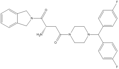
Chemical_Nomenclature : (2S)-2-amino-4-[4-[bis(4-fluorophenyl)methyl]piperazin-1-yl]-1-(1,3-dihydroisoindol-2-yl)butane-1,4-dione
Canonical SMILES : C1CN(CCN1C(C2=CC=C(C=C2)F)C3=CC=C(C=C3)F)C(=O)CC(C(=O)N4CC5=CC=CC=C5C4)N
InChI : InChI=1S\/C29H30F2N4O2\/c30-24-9-5-20(6-10-24)28(21-7-11-25(31)12-8-21)34-15-13-33(14-16-34)27(36)17-26(32)29(37)35-18-22-3-1-2-4-23(22)19-35\/h1-12,26,28H,13-19,32H2\/t26-\/m0\/s1
InChIKey : ZKIQFLSGMMYCGS-SANMLTNESA-N
Other name(s) : CHEMBL1814633,GTPL8551,C29H30N4O2F2,MolPort-035-706-578,BDBM50350169
MW : 504.57
Formula : C29H30F2N4O2
CAS_number :
PubChem : 56658139
UniChem : ZKIQFLSGMMYCGS-SANMLTNESA-N
IUPHAR : 8551
Wikipedia :
Target
Families : 1G244 ligand of proteins in family: DPP4N_Peptidase_S9
Stucture :
Protein : human-DPP8 || human-DPP9
References (9)
| Title : The antiatherosclerotic action of 1G244 - An inhibitor of dipeptidyl peptidases 8\/9 - is mediated by the induction of macrophage death - Wisniewska_2023_Eur.J.Pharmacol__175566 |
| Author(s) : Wisniewska A , Czepiel K , Stachowicz A , Pomierny B , Kus K , Kiepura A , Stachyra K , Surmiak M , Madej J , Olszanecki R , Suski M |
| Ref : European Journal of Pharmacology , :175566 , 2023 |
| Abstract : Wisniewska_2023_Eur.J.Pharmacol__175566 |
| ESTHER : Wisniewska_2023_Eur.J.Pharmacol__175566 |
| PubMedSearch : Wisniewska_2023_Eur.J.Pharmacol__175566 |
| PubMedID: 36739078 |
| Title : Non-Specific Inhibition of Dipeptidyl Peptidases 8\/9 by Dipeptidyl Peptidase 4 Inhibitors Negatively Affects Mesenchymal Stem Cell Differentiation - Torrecillas-Baena_2023_J.Clin.Med_12_4632 |
| Author(s) : Torrecillas-Baena B , Camacho-Cardenosa M , Quesada-Gomez JM , Moreno-Moreno P , Dorado G , Galvez-Moreno M , Casado-Diaz A |
| Ref : J Clin Med , 12 :4632 , 2023 |
| Abstract : Torrecillas-Baena_2023_J.Clin.Med_12_4632 |
| ESTHER : Torrecillas-Baena_2023_J.Clin.Med_12_4632 |
| PubMedSearch : Torrecillas-Baena_2023_J.Clin.Med_12_4632 |
| PubMedID: 37510747 |
| Gene_locus related to this paper: human-DPP8 , human-DPP9 |
| Title : Aerosol-based ligand soaking of reservoir-free protein crystals - Ross_2021_J.Appl.Crystallogr_54_895 |
| Author(s) : Ross B , Krapp S , Geiss-Friedlander R , Littmann W , Huber R , Kiefersauer R |
| Ref : J Appl Crystallogr , 54 :895 , 2021 |
| Abstract : Ross_2021_J.Appl.Crystallogr_54_895 |
| ESTHER : Ross_2021_J.Appl.Crystallogr_54_895 |
| PubMedSearch : Ross_2021_J.Appl.Crystallogr_54_895 |
| PubMedID: 34188616 |
| Gene_locus related to this paper: human-DPP8 |
| Title : Decrease of the pro-inflammatory M1-like response by inhibition of dipeptidyl peptidases 8\/9 in THP-1 macrophages - quantitative proteomics of the proteome and secretome - Suski_2020_Mol.Immunol_127_193 |
| Author(s) : Suski M , Wisniewska A , Kus K , Kiepura A , Stachowicz A , Stachyra K , Czepiel K , Madej J , Olszanecki R |
| Ref : Mol Immunol , 127 :193 , 2020 |
| Abstract : Suski_2020_Mol.Immunol_127_193 |
| ESTHER : Suski_2020_Mol.Immunol_127_193 |
| PubMedSearch : Suski_2020_Mol.Immunol_127_193 |
| PubMedID: 32998073 |
| Title : DPP8 is a novel therapeutic target for multiple myeloma - Sato_2019_Sci.Rep_9_18094 |
| Author(s) : Sato T , Tatekoshi A , Takada K , Iyama S , Kamihara Y , Jawaid P , Rehman MU , Noguchi K , Kondo T , Kajikawa S , Arita K , Wada A , Murakami J , Arai M , Yasuda I , Dang NH , Hatano R , Iwao N , Ohnuma K , Morimoto C |
| Ref : Sci Rep , 9 :18094 , 2019 |
| Abstract : Sato_2019_Sci.Rep_9_18094 |
| ESTHER : Sato_2019_Sci.Rep_9_18094 |
| PubMedSearch : Sato_2019_Sci.Rep_9_18094 |
| PubMedID: 31792328 |
| Gene_locus related to this paper: human-DPP8 |
| Title : Dipeptidyl peptidase 9 (DPP9) in human skin cells - Gabrilovac_2017_Immunobiology_222_327 |
| Author(s) : Gabrilovac J , Cupic B , Zapletal E , Kraus O , Jakic-Razumovic J |
| Ref : Immunobiology , 222 :327 , 2017 |
| Abstract : Gabrilovac_2017_Immunobiology_222_327 |
| ESTHER : Gabrilovac_2017_Immunobiology_222_327 |
| PubMedSearch : Gabrilovac_2017_Immunobiology_222_327 |
| PubMedID: 27682012 |
| Gene_locus related to this paper: human-DPP9 |
| Title : Inhibition of dipeptidyl peptidase 8\/9 impairs preadipocyte differentiation - Han_2015_Sci.Rep_5_12348 |
| Author(s) : Han R , Wang X , Bachovchin W , Zukowska Z , Osborn JW |
| Ref : Sci Rep , 5 :12348 , 2015 |
| Abstract : Han_2015_Sci.Rep_5_12348 |
| ESTHER : Han_2015_Sci.Rep_5_12348 |
| PubMedSearch : Han_2015_Sci.Rep_5_12348 |
| PubMedID: 26242871 |
| Title : Dipeptidyl peptidases in atherosclerosis: expression and role in macrophage differentiation, activation and apoptosis - Matheeussen_2013_Basic.Res.Cardiol_108_350 |
| Author(s) : Matheeussen V , Waumans Y , Martinet W , Van Goethem S , Van der Veken P , Scharpe S , Augustyns K , De Meyer GR , De Meester I |
| Ref : Basic Res Cardiol , 108 :350 , 2013 |
| Abstract : Matheeussen_2013_Basic.Res.Cardiol_108_350 |
| ESTHER : Matheeussen_2013_Basic.Res.Cardiol_108_350 |
| PubMedSearch : Matheeussen_2013_Basic.Res.Cardiol_108_350 |
| PubMedID: 23608773 |
| Title : Biochemistry, pharmacokinetics, and toxicology of a potent and selective DPP8\/9 inhibitor - Wu_2009_Biochem.Pharmacol_78_203 |
| Author(s) : Wu JJ , Tang HK , Yeh TK , Chen CM , Shy HS , Chu YR , Chien CH , Tsai TY , Huang YC , Huang YL , Huang CH , Tseng HY , Jiaang WT , Chao YS , Chen X |
| Ref : Biochemical Pharmacology , 78 :203 , 2009 |
| Abstract : Wu_2009_Biochem.Pharmacol_78_203 |
| ESTHER : Wu_2009_Biochem.Pharmacol_78_203 |
| PubMedSearch : Wu_2009_Biochem.Pharmacol_78_203 |
| PubMedID: 19439267 |